
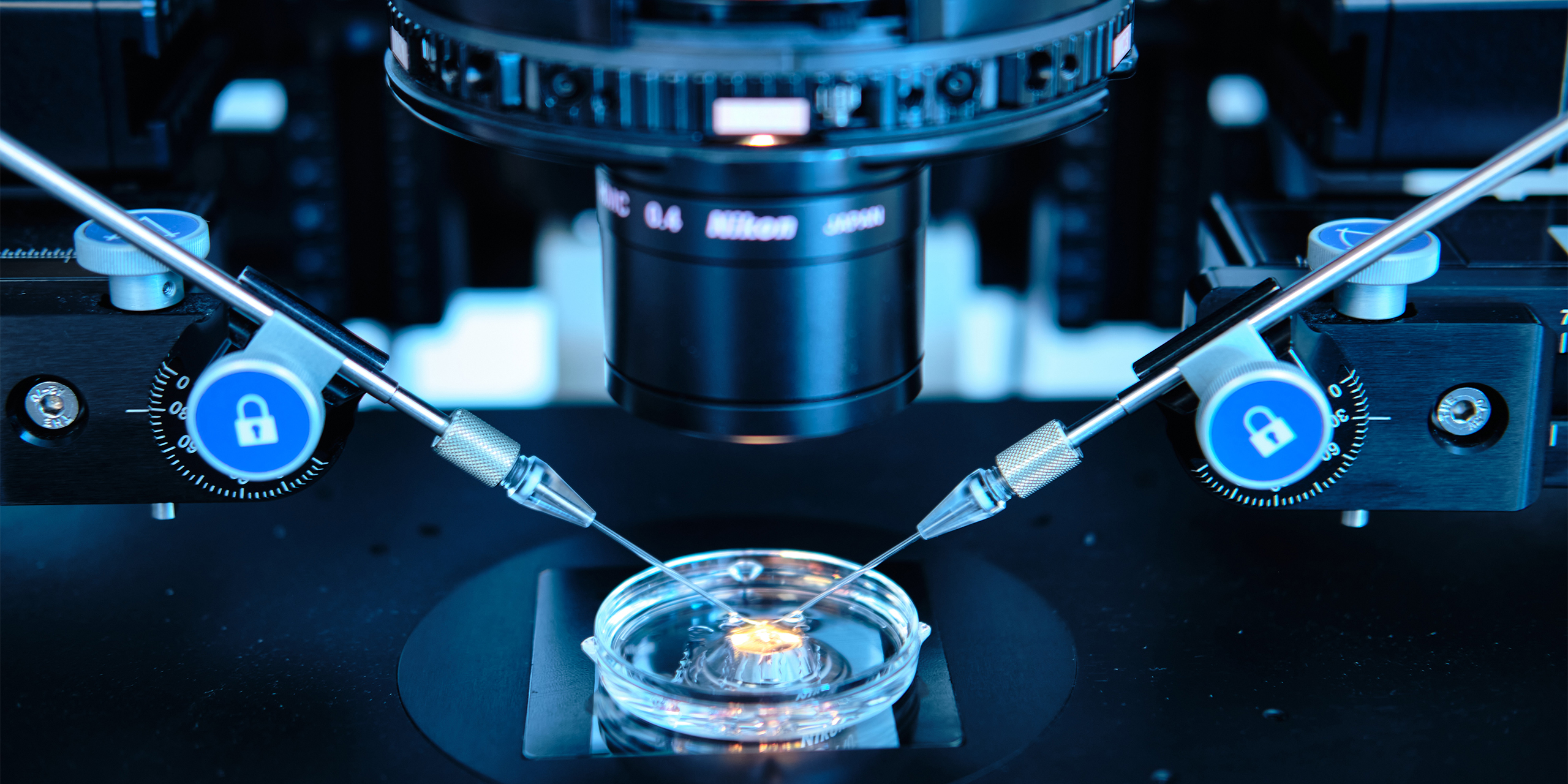
Unwanted childlessness is an important topic worldwide and different reasons apart from sub- and infertility led to this trend. On the female side, the increasing age leads to a significant decrease of becoming pregnant naturally and reduces the effectiveness of possible treatments to overcome sub- and infertility. Since the beginning of the records of the German IVF registry (1997), 319.119 children have been born using assisted reproduction in Germany [1] and since more than four decades fertilization and early embryo development can also take place artificially in a petri dish. This means that eggs, sperm, and embryos are handled outside of their natural environment in an artificial environment for a period of time that ranges from hours to days. The environment should be as similar as possible to the natural surrounding. To further improve the quality of embryo culture media and to mimic in vivo nutrition, growth factors like granulocyte macrophage colony-stimulating factor (GM-CSF) became attractive candidates for medium supplementation. GM-CSF is a cytokine influencing the maternal-fetal interface and supporting placental development in mouse and human. It is expressed in epithelial cells of the endometrium under regulation of estrogens. A large clinical trial showed that GM-CSF supplemented embryo culture medium leads to an increased survival of embryos up to week 12 [2]. Especially in women with previous miscarriages GM-CSF supplementation of culture media improved implantation rates. Animal and cell culture studies on trophectoderm cells supported an effect on cellular expansion and pluripotency pathways after exposure to GM-CSF [3].
Literature:
[1] Deutsches IVF-Register 2019 (D·I·R); Journal für Reproduktionsmedizin und Endokrinologie
[2] Ziebe S, Loft A, Povlsen BB, Erb K, Agerholm I, Aasted M, Gabrielsen A, Hnida C, Zobel DP, Munding B, Bendz SH, Robertson SA. A randomized clinical trial to evaluate the effect of granulocyte-macrophage colony-stimulating factor (GM-CSF) in embryo culture medium for in vitro fertilization. Fertil Steril. 2013 May;99(6):1600-9. doi: 10.1016/j.fertnstert.2012.12.043.
[3] Sjöblom C, Wikland M, Robertson SA. Granulocyte-macrophage colony-stimulating factor (GM-CSF) acts independently of the beta common subunit of the GM-CSF receptor to prevent inner cell mass apoptosis in human embryos. Biol Reprod. 2002 Dec;67(6):1817-23. doi: 10.1095/biolreprod.101.001503.
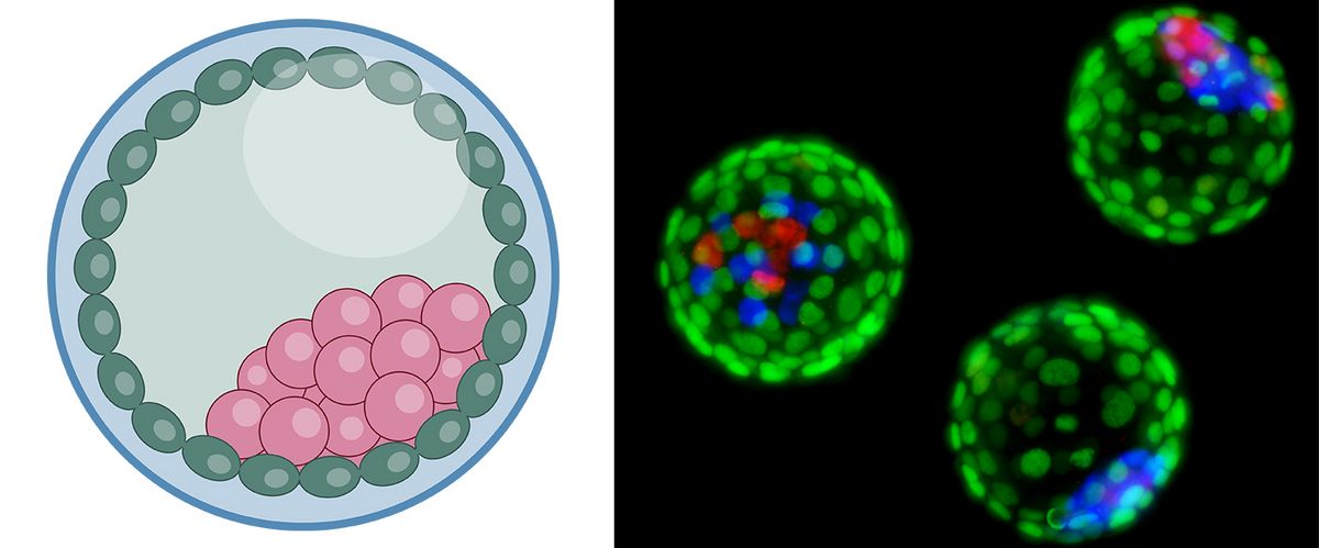
The success of ART is a healthy live birth. To reach this goal researchers and clinicians try to increase pregnancy rates by supporting the pre-implantation embryo as good as possible. However, it is not only important to support the embryo at its best, but it is also equally important to do no harm to the embryo. This holds true also for a culture medium that should have no influence on the normal developmental program of an embryo. Aim of this study is to investigate, if the supplementation of GM-CSF in a human ART medium or in a mouse optimized medium leads to a change in cell number and cell lineages in the early pre-implantation mouse embryo.
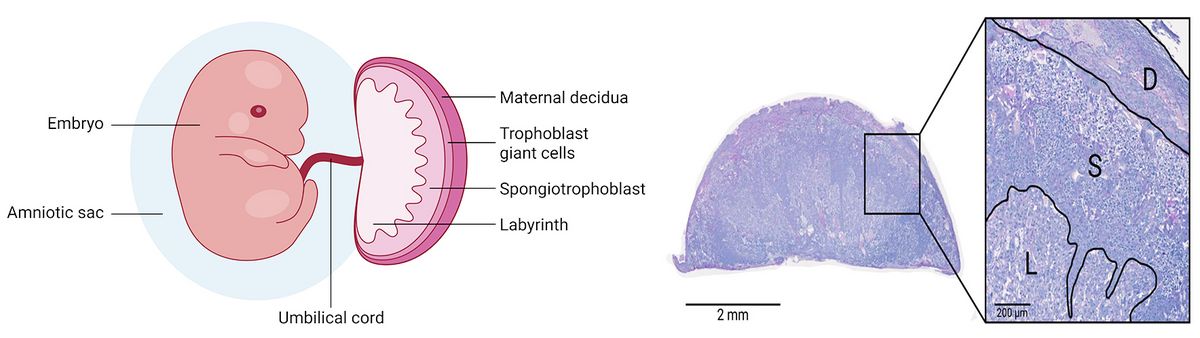
In this study we hypothesize that different concentrations of GM-CSF supplemented into a culture medium have an effect on trophectoderm cells which then affects placental development during and after implantation. The aim of this study is to examine if an in vitro preimplantation cultivation with GM-CSF has any effect on the morphological structure of the placenta.
Michele Boiani, Max Planck Institute for Molecular Biomedicine, Münster Website
Séverine le Gac, MESA+, Enschede Website Séverine le Gac Website MESA+
Anika Witten, Department Genetic Epidemiology at the Institute for Human Genetics, Münster Website
Georg Fuellen, Institute for Biostatistics and Informatics in Medicine and Ageing Research, Rostock Website