News
Das "Paper of the Month" 10/2022 geht an: AG Rescher aus dem Institut für Medizinische Biochemie
Für den Monat Oktober 2022 geht das „Paper of the Month“ der Medizinischen Fakultät der WWU Münster an die:
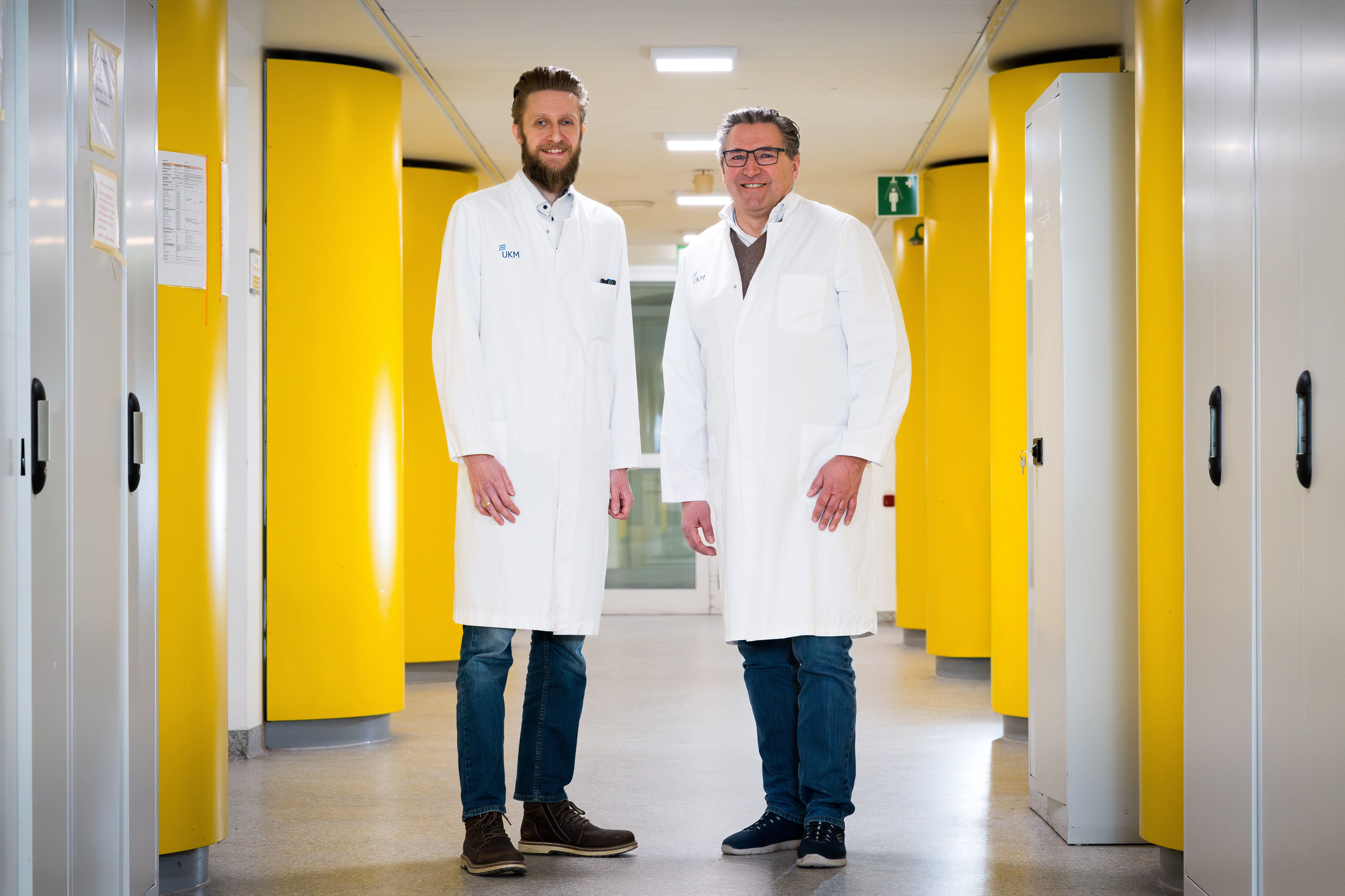
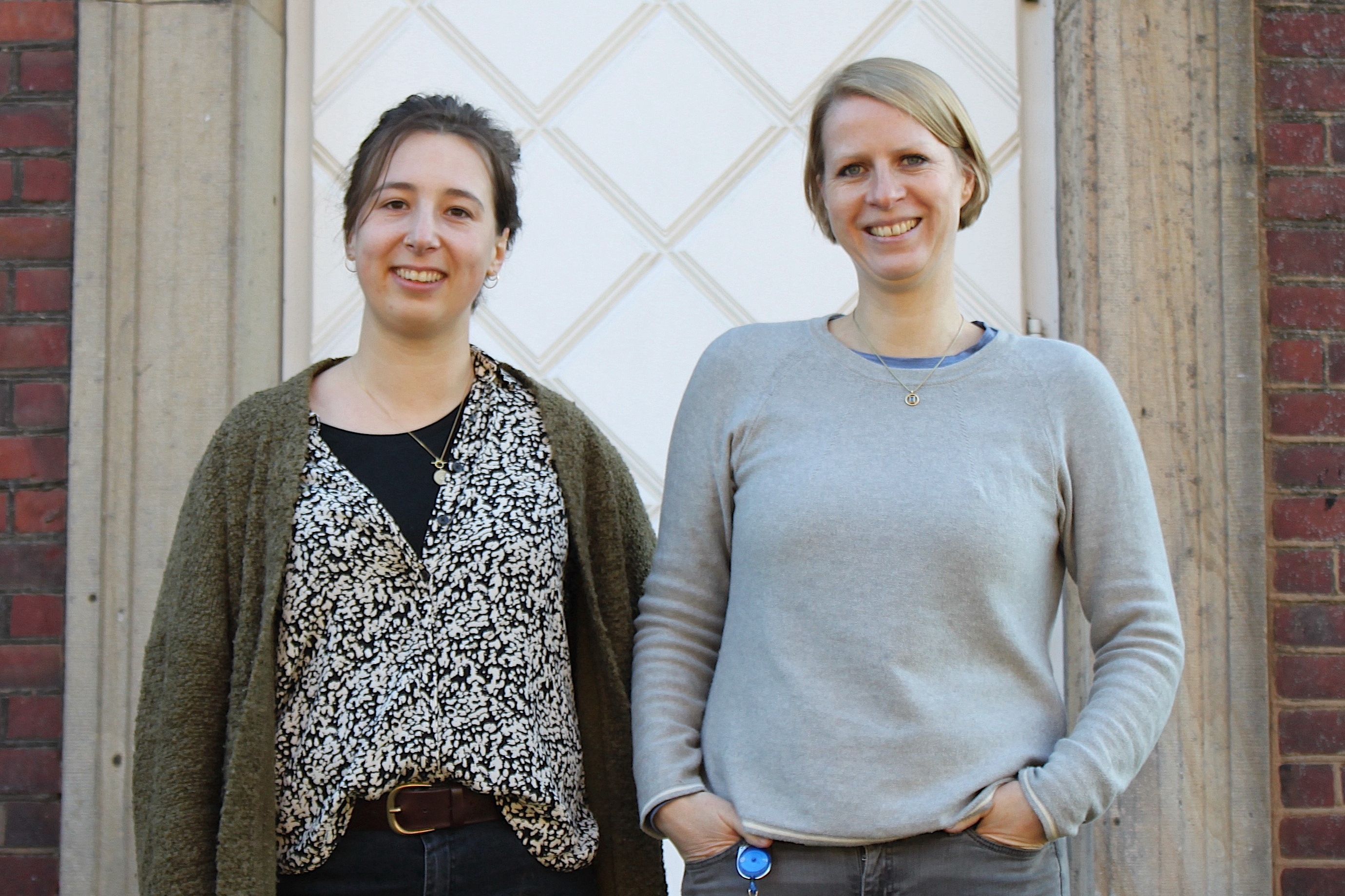
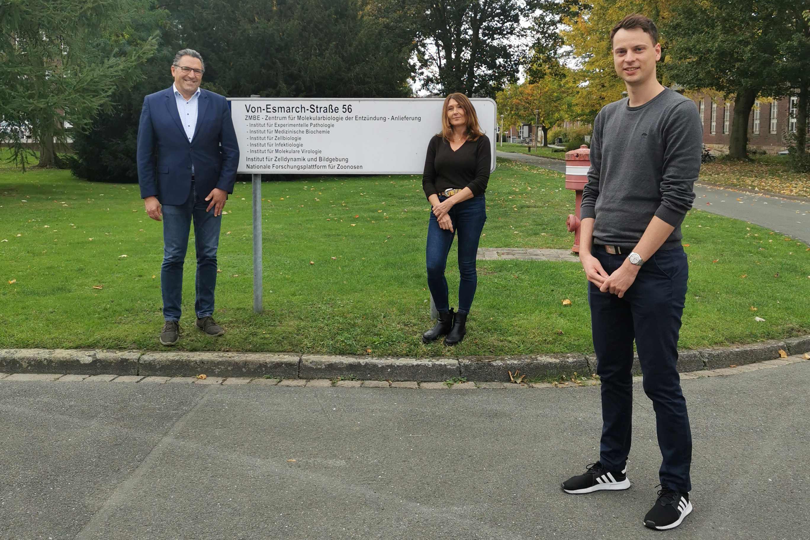
Arbeitsgruppe Prof. Rescher aus dem Institut für Medizinische Biochemie
Schloer, S; Treuherz, D; ...; Brunotte, L; Rescher, U.
SEP 2022 | EMERGING MICROBES & INFECTIONS
Begründung der Auswahl:
In dieser Publikation wird die Etablierung eines 3D-ex-vivo-Gewebemodells zur Untersuchung der Pathophysiologie einer viralen Infektion im frühen Stadium beschrieben. Dieses ist ein sehr hilfreiches und fortschrittliches Tool: zum einen für die hochrelevante virologische Forschung selbst, aber auch, um die human-transnationale Forschung voranzubringen, Tierversuche zu reduzieren und Drug-Screenings durchzuführen.
Zu Hintergrund, Fragestellung und Bedeutung der Publikation:
Viruspandemien bedrohen die Gesundheit und die weltweite Wirtschaft. Zellkulturbasierte Modelle des viralen Lebenszyklus spiegeln nicht die physiologische Umgebung wider, Tiermodelle sind aber ethisch umstritten. Zur Untersuchung von Virus-Wirt-Interaktionen in einem natürlichen Wirt haben wir neue ex-vivo-Infektionsmodelle aus tierischen und humanem Lungengewebe entwickelt und verglichen.
Die auf Lungengewebeexplantaten basierenden ex-vivo-Infektionsmodelle unterstützten eine effiziente Virusreplikation von Influenza A und SARS-CoV-2, setzten entzündlicher Zytokine frei und rekapitulierten den Beginn der Interferonantwort, einem bekannten zellulären Programm zur Induktion der Abwehr von Pathogenen. Die Tiermodelle waren vergleichbar mit ex-vivo-Infektionen in menschlichen Lungenexplantaten. Manipulationen der Wirts-Endolysosomen störten die Virus-Endosomen-Fusion, verringerten die Virustiter und die Anzahl infizierter Zellen und korrelierten mit einer reduzierten Immunantwort. Unsere Modelle bestätigen, dass die endolysosomale Wirt-Pathogen-Schnittstelle durch die Umwidmung klinisch zugelassener Medikamente gezielt für pharmakologische Interventionen eingesetzt werden kann.
Unsere ex-vivo-Lungenmodelle können in einem Standard-Zellkulturlabor ohne ausgeklügelte Ausrüstung aufgebaut werden, eignen sich als präklinische Plattformen, um die Anfangsphase der viralen Atemwegsinfektion in einem physiologisch sinnvollen Umfeld zu rekapitulieren und können als praktisches Modell für Medikamententests verwendet werden.
Förderhinweise: siehe unten, im englischen Text
Background and fundamental question of the publication:
Viral pandemics pose massive threats to global health and economics. Cell culture-based models to study the viral life cycle do not reflect the physiologic environment, yet animal models are linked with ethical questions. To bridge the gap between cell culture and animal models for studying virus-host interactions in a natural host, we established and compared the suitability of novel murine, hamster, and human lung tissue explant platforms.
Both the animal and the human ex vivo infection models based on lung tissue explants sustained efficient influenza A and SARS-CoV-2 viral replication, the release of inflammatory cytokines, and recapitulated the onset of the interferon response, a well-known antimicrobial program to induce cellular antimicrobial and particularly antiviral defense. The animal models were comparable to ex vivo infection in human lung explants. Manipulations of the host endolysosomes interfered with virus-endosome fusion. The marked reduction in virus titres and numbers of infected cells correlated with a reduced immune response, thus confirming that the endolysosomal host-pathogen interface can be targeted for pharmacological intervention via the repurposing of clinically licensed drugs.
Our ex vivo lung models can be set up in a standard cell culture lab at the required safety level without sophisticated equipment, are suited as preclinical platforms to recapitulate the initial phase of viral airway infection in a physiologically meaningful setting, and can be used as a convenient model for drug testing.
This research was funded by grants from the German Research Foundation (DFG) CRC1009 “Breaking Barriers”, Project A06 (to U.R.) and B02/B1302 (to S.L.), CRC 1348 “Dynamic Cellular Interfaces”, Project A11 (to U.R.), DFG Lu477/23-21 (to S.L.), KFO342 P6 (to L.B. and S.L.), the Interdisciplinary Center for Clinical Research (IZKF) of the Münster Medical School, grant numbers Re2/022/20 (to U.R.) and Lud4/013/21 (to S.L) and Bru2/015/19 (to L.B.) as well as funding of the Innovative Medizinische Forschung (IMF) of the medical faculty of the University Muenster, grant number BR111905 (to L.B.) and SC121912 (to S.S.). Furthermore, funding was provided by the Federal Ministry of Education and Research, grant 01KI20218 (Co-Immune) to L.B. and 01KX2021 (NUM-COVID-19, Organo-Strat) to L.B. and S.L. and by the Bundesministerium für Gesundheit ZMI1-2519PAT701 (to S.K.).
Die bisherigen ausgezeichneten „Papers of the Month“ finden Sie HIER.


