
Magnetic Resonance Imaging
Magnetic resonance imaging (MRI), also known as nuclear magnetic resonance imaging, is an imaging technique used, among other applications, in medical diagnostics to visualize the structure and function of tissues and organs within the body. It is based on very strong magnetic fields and electromagnetic alternating fields in the radiofrequency range, which are used to resonantly excite specific atomic nuclei (most commonly hydrogen nuclei) in the body. These nuclei then induce electrical signals in a receiver circuit.
MRI enables the generation of cross-sectional images of the human (or animal) body, allowing assessment of organs and many pathological changes. A key factor underlying image contrast is the difference in relaxation times between various tissue types. In addition, variations in hydrogen atom content across different tissues (e.g., muscle, bone, etc.) also contribute to image contrast.
MRI provides excellent soft-tissue contrast. This results from differences in signal intensity originating from various soft tissues. The technique does not involve potentially harmful ionizing radiation. Furthermore, by adjusting imaging parameters, a very high level of detail can be achieved, surpassing that of X-ray imaging or computed tomography (CT).
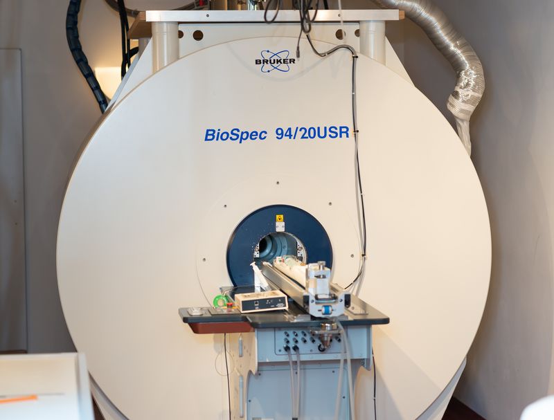
For our projects, we work with clinical 1.5 T and 3 T systems, as well as a preclinical 9.4 T system (Experimental Magnetic Resonance Group).