
Optical Imaging
Optical imaging, also known as fluorescence imaging, is based on the visualization of a substance that fluoresces in the near-infrared range within the organism. Light in the near-infrared spectrum (approximately 600–950 nm), unlike visible light, can penetrate tissue to a depth of several centimeters. These substances can therefore be imaged even deep within the body using photographic or microscopic techniques.
When coupled to specific probes, these dyes can be used to visualize and further characterize disease sites or particular cell populations. The migration of individual labeled cells can also be made visible using optical imaging methods (cell tracking).
Fluorescence Reflectance Imaging – fluorescence-based imaging
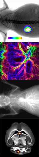
Fluorescence Reflectance Imaging (FRI), analogous to conventional planar X-ray imaging or classical photography, is a projection-based technique in which signals from different tissue depths are mapped onto a two-dimensional plane (at the level of the body surface). Due to scattering and absorption of fluorescence from deeper tissue layers, depth resolution is therefore very limited, and quantitative analysis of the results is restricted.
To nevertheless achieve an approximate anatomical localization of detected signals, white-light and X-ray images are used, which can then be overlaid with the fluorescence images.