

Newly discovered mechanism of primary ciliary dyskinesia: Researchers identify defects in a previously unknown ciliary linker (Transition zone linker) at the ciliary control gate as a previously overlooked cause of the disease
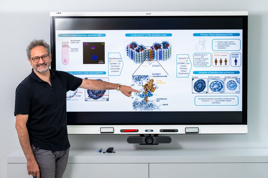
Successful teamwork: Professor Heymut Omran points to the transition zone linkers. His international research team is the first to show that defects in these structures can cause PCD. (Photo: University of Münster/Michael Ibrahim).
Münster/Boston – An international research team from the University of Münster’s Faculty of Medicine, led by Prof. Dr. Heymut Omran, and Harvard Medical School in Boston, led by Prof. Dr. Alan Brown, has elucidated a previously unknown molecular mechanism underlying the rare genetic disorder primary ciliary dyskinesia (PCD).
When the lungs’ “cleaning crew” goes on strike
Motile cilia line the airways and transport mucus, pathogens, and particles out of the lungs, forming a first line of defense that prevents infections and keeps the airways clear. In PCD, this cleaning mechanism is disrupted: ciliary transport is impaired, mucus accumulates, and those affected suffer from chronic respiratory infections and progressive lung damage.
Most of the PCD-associated genes known to date encode proteins of the biological motor complexes that generate or control ciliary beating. However, in approximately 25% of clinically diagnosed patients, no causative mutation is found in any of these known genes, suggesting that important components of the cilia remain undiscovered.
The transition zone as a control gate for the cilia
The research team suspected that the so-called transition zone - a region at the base of each cilium - might play a role that had been underestimated until now. This structure functions like a control gate that regulates which proteins enter and exit the cilia.
A high-resolution look inside the cell
To determine the molecular structure of the transition zone in human motile cilia for the first time, the team used state-of-the-art imaging techniques, in particular in situ cryo-electron tomography on human airway epithelial cells. In the process, they identified a previously unknown multiprotein complex (linker) consisting of four components (ECT2L, SKP1, DZANK1, and DYNLL1/2) that connects adjacent microtubules. Using immunofluorescence and expansion microscopy, the team also demonstrated that many of these proteins occur specifically, and in some cases exclusively, in multiciliated epithelial cells and are specifically localized to the transition zone of motile cilia.
The genetic evidence: Four patients, one common defect
While analyzing approximately 500 individuals with suspected PCD, the team identified mutations in the ECT2L and DZANK1 genes in four patients. These patients exhibited the typical clinical features of PCD: chronic productive cough, recurrent sinusitis, recurrent pneumonia, and bronchiectasis. Respiratory tract cells from these patients exhibited a disrupted transition zone, abnormally swollen cilia tips (so-called “hockey-stick cilia”), and impaired clearance function. Knocking out ECT2L in a cell culture model precisely reproduced these structural and functional defects.
Implications for research
“These findings provide, for the first time, direct genetic evidence that defects in transition zone linkers can lead to diseases of motile cilia in humans,” explains Prof. Heymut Omran. “They also demonstrate how structural biology methods can uncover new disease mechanisms directly in biological source material and provide a promising explanatory framework for some of the previously unexplained cases of PCD.” Since these structures are evolutionarily conserved from single-celled organisms to humans, these findings are also of great significance for the entire field of biology.
The study establishes the transition zone linker as an essential element for the function of motile cilia and was published in the prestigious journal Science.
Link to the study: https://www.science.org/doi/10.1126/science.aei5957