Molecular basis of congenital laterality defects, including heart and vascular malformations
Our group is conducting a study to investigate the causes of lateralization defects (abnormal location and function of organs). Among congenital malformations, heart defects and abnormalities of the vessels near the heart are the most commonly represented (approximately 0.8% of all newborns are affected). These disorders require diagnosis and intervention (invasive cardiac surgery) at infancy. Some patients with congenital heart defects may also suffer from dysfunction of other organ systems or positional anomalies of the heart and internal organs. The aim of our study is to find the responsible genes for these disorders and understand their function in laterality defects, including congenital heart and vascular malformations. In half of all patients with primary ciliary dyskinesia (PCD), the arrangement of the left-right body axis is abnormal. In most cases, this results in situs inversustotalis, which is the complete mirror-image arrangement of internal organs. In approximately 6% of patients, the organs are positioned in a way that neither the normal arrangement (situs solitus) or complete reversal (situs inversus) can be determined. This condition is known as situs ambiguus. In contrast to situs inversus, situs ambiguus is often associated with a malformation or absence of an organ. Left isomerism is often associated with polysplenia, whereas right isomerism usually lacks the facility of the spleen (asplenia). In 3% of cases, heterotaxy is associated with a congenital heart defect.

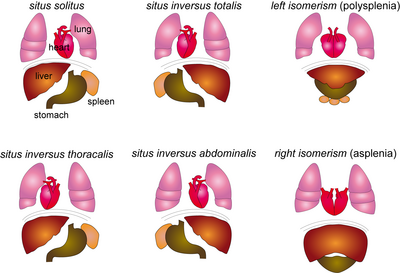
Figure 1: Schematic of possible Situs variations. In situs ambiguous, four laterality defects can be distinguished: thoracic situs inversus, abdominal situs inversus, and left or right isomerism. In both thoracic and abdominal situs inversus, only part of the organs are abnormally arranged – organs above the diaphragm for thoracic situs inversus and organs below the diaphragm for situs inversus abdominalis. For isomerism, the affected side (left or right) is a mirror image of the affected organs. However, even combinations of these four categories are possible.
The first results of our study suggest that patients with heart malformations harbor mutations in a gene that is causative for PCD. The severity of heart disease overshadows the symptoms of respiratory disease in these patients, thus we would like to examine a larger cohort of patients with situs anomalies for the presence of PCD. For this we need EDTA or heparin-treated blood from patients and, if possible, their families. The submission of brush biopsy specimens allows us to quickly screen for defects of the cilia of respiratory epithelial cells, which are present in most of the PCD patients.
If you wish to support our study as a patient or doctor, please find the relevant documents and additional information in the following links:

