
APOL1 as a risk factor for kidney diseases
APOL1 as a risk factor for kidney diseases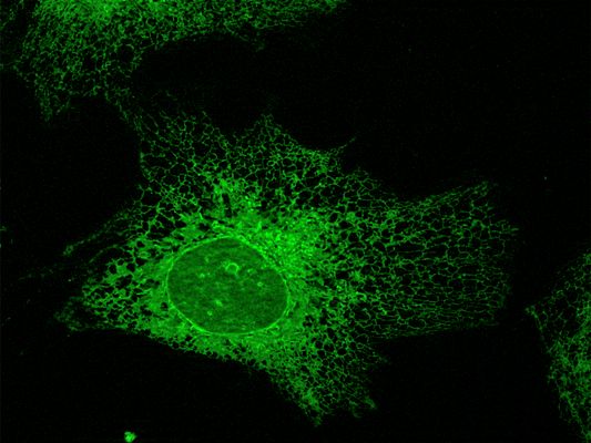
Subspecies of the single-celled parasite Trypanosoma brucei cause African sleeping sickness (also known as African trypanosomiasis). This tropical disease is transmitted by the tsetse fly and occurs primarily in various tropical regions of Africa. Human blood serum contains a factor that normally protects people from this disease. In 2003, this factor was identified as APOL1 (short for Apolipoprotein L1). APOL1 — which occurs only in humans and a few other higher primates — is both necessary and sufficient to trigger this trypanolytic effect.
A co-evolutionary process between the SRA resistance gene (serum resistance-associated protein) of the trypanosomes and the trypanolytic factor APOL1 has led to the emergence of two APOL1 gene variants (G1 and G2), which are relatively common among African Americans and populations in West Africa.
In 2010, two research groups independently demonstrated that these specific APOL1 variants are associated with an increased risk of HIV-associated nephropathy (HIVAN). Subsequent studies have repeatedly confirmed this link. At the same time, it became evident that individuals who are homozygous for these APOL1 variants also carry a higher risk for other kidney diseases. Kidney diseases associated with APOL1 are also referred to as APOL1-mediated kidney diseases (AMKD).
However, not every carrier of APOL1 risk variants develops disease. It is therefore assumed that in addition to the genetic predisposition, at least one other factor (a "second hit") is necessary to trigger kidney disease. Elevated cytokine concentrations — such as interferons — or viral infections (particularly HIV or COVID-19) may play a particularly important role in this context.
Research on APOL1 thus lies at the intersection of parasitology, evolutionary biology, infectious diseases, and nephrology. On a molecular level, however, it remains largely unclear what exact function APOL1 normally serves in the cell, and how it differs from other members of the APOL gene family. In previous work, we were able to show that APOL1 is primarily localized to the endoplasmic reticulum (ER), and that increased expression of the APOL1 risk variants leads to energy depletion in cells.
In our project funded by the German Research Foundation (DFG) ( WE 2550/3-1 & 3-2; project number 324783603), we are now using primarily cell biological approaches to more precisely analyze the impact of APOL1 on ER-associated functions (e.g., biosynthesis, protein secretion).
The role of the Crumbs2 protein in podocytes
The role of the Crumbs2 protein in podocytesCell polarity refers to the ordered but asymmetric distribution of proteins and membrane components within a cell. It forms the basis for many essential cellular processes. Both cell polarization and the associated formation of cell–cell junctions (tight junctions and adherens junctions) are regulated by evolutionarily conserved multiprotein complexes.
One such complex is the Crumbs complex, which consists of the core components Crumbs, Pals1 (see Project Pals1), and Patj, and primarily regulates the establishment of apical membranes within the plasma membrane.
In humans, there are three different Crumbs genes (CRB1, CRB2, and CRB3). All Crumbs proteins share a common structure: an N-terminal extracellular domain (ECD), a transmembrane domain (TMD), and a C-terminal intracellular domain (ICD). The most significant differences lie in the ECD. In CRB1 and CRB2, the ECD is large and contains several EGF-like and Laminin G domains, while the much smaller CRB3 proteins lack most of the ECD.
In the kidney, CRB2 and two splice variants of CRB3 — CRB3A and CRB3B — are expressed. Both CRB3 isoforms are found in all renal epithelial cells, whereas CRB2 is primarily expressed in glomerular podocytes. These are highly polarized, post-mitotic cells that form part of the glomerular filtration barrier. Between their extensively branched cellular processes, podocytes form unique cell–cell junctions, known as slit diaphragms, which are exclusive to this cell type.
Recently, it was shown that specific mutations in the human CRB2 gene are associated with severe damage to the renal filtration barrier. This suggests that CRB2 has podocyte-specific functions that are essential for maintaining the integrity of this barrier.
The goal of our DFG-funded project (DFG WE 2550/2-2; project number 469263956) is to analyze in detail the cell biological functions of CRB2 and their significance for podocytes. To achieve this, we use various cell systems in combination with advanced microscopy techniques. The project is conducted in close collaboration with the research group of Prof. Ulrich Kubitscheck (Biophysikalische Chemie, University of Bonn).
The role of Pals1 in signal transduction processes in epithelia
The role of Pals1 in signal transduction processes in epitheliaThe mammalian kidney consists of approximately 1–2 million functional units known as nephrons, which can be broadly divided into a glomerulus and a tubule. Blood serum filtration takes place in the glomeruli, whereas the reabsorption of essential biomolecules (e.g., water, salts, sugars, amino acids, etc.) and the associated concentration of primary urine are primarily carried out by the tubular epithelia of the nephron — also referred to as segments.
Although the epithelial cells of different nephron segments vary significantly in their morphology and physiology, they all exhibit pronounced apico-basal cell polarity.
In previous work, we analyzed the role of Pals1 in various renal epithelia. Pals1 (gene: MPP5) interacts with all components of the Crumbs complex through its multiple domains and also mediates interactions between the Crumbs complex and another polarity complex — the aPKC complex.
We were able to show that mice with reduced Pals1 expression in the nephron develop polycystic kidneys and severe proteinuria, indicating defects in tubular epithelia and a compromised renal filtration barrier. These severe impairments are accompanied by alterations in Hippo and TGFβ signaling pathways. This suggests that Pals1 is not only structurally important but also serves as a crucial hub for signal transduction.
This DFG-funded project (DFG WE2550/4-1; project number 414057425) is conducted in close collaboration with the research group of Prof. Michael Krahn (Medical Cell Biology, MedD, University of Münster) and is also part of the DFG Priority Programme “Epithelial Intercellular Junctions as Dynamic Hubs to Integrate Forces, Signals, and Cell Behaviour” (SPP1782).