The focus of our research is the adaptation and development of high-resolution imaging techniques for studying cellular signaling and trafficking, with the emphasis on
presynaptic mechanisms during synaptic transmission. At the synapse, neurotransmitter is rapidly released from small vesicles which are triggered to fuse with the plasma membrane by the entry of Ca2+ ions (Fig.1).
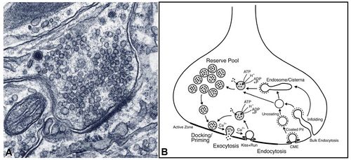
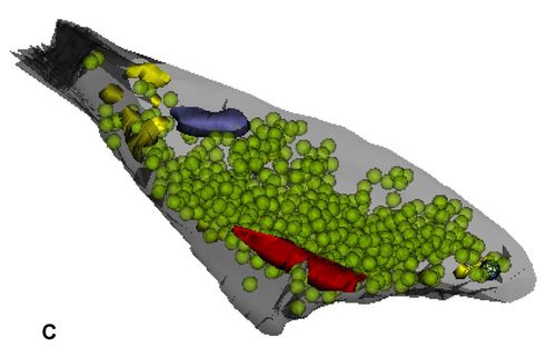
Fig.1 A: A synapse from rat hippocampus after 14 days in culture as seen under the electron microscope. It contains some 100 – 200 synaptic vesicles filled with neurotransmitter, some of which are docked at the ‘active zone’ (black thickening of the membrane) and ready to fuse upon electric stimulation. In B different putative mechanisms of vesicle recycling are schematically depicted. C: 3D reconstruction of a hippocampal synapse from serial EM sections (red: active zone; green: vesicles; blue: mitochondria; yellow: endocytic intermediates).
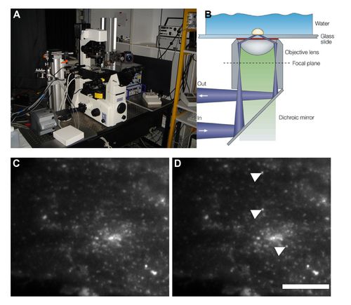
The maintenance of synaptic transmission requires that these vesicles are retrieved by a reverse process, endocytosis. To delineate the mechanisms by which synaptic vesicle components can be retrieved we employ high- resolution imaging techniques, like two-photon laser scanning microscopy, fluorescence photo-activation localization microscopy (FPALM), and total internal reflection microscopy (TIRFM, Fig.2), as well as electrophysiology, electron microscopy including 3D reconstruction (Fig.1), and molecular biology approaches.
Fig2: A,B: Setup and principle of total internal reflection microscopy (TIRFM) C,D: TIRF time lapse images of a fibroblast footprint illustrating exocytosis of single vesicles (marked by arrowheads). Vesicles are endogenously labelled with a pH sensitive variant of green fluorescent protein (pHluorin) fused to transferrin receptors which are cycled between plasma membrane and the acidic endosomes (scale bar : 5 μm).
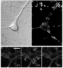
By overexpression of endogenous fluorescent probes (e.g. synaptopHluorin Fig.3) and the usage of knock-out models we can target or modulate specific proteins thought to be pivotal in synaptic vesicle endocytosis.
Fig.3: A,B: DIC and fluorescence images of hippocampal neurons in culture. C: Synaptic vesicles at single active synaptic boutons are labeled with the activity-dependent fluorescent marker FM1-43. C: Fluorescence signals from single fusing vesicles at synaptopHluorin-expressing boutons (marked by arrowheads, scale bar 5 µm).
Selected Publications
Schmidt NC, Kahms M, Hüve J, Klingauf J. (2018) Intrinsic refractive index matched 3D dSTORM with two objectives: Comparison of detection techniques. Sci. Rep. 8(1):13343.
Martineau M, Guzman RE, Fahlke C, Klingauf J. (2017) VGLUT1 functions as a glutamate/proton exchanger with chloride channel activity in hippocampal glutamatergic synapses. Nat. Commun. 8(1):2279.
Bodzęta A, Kahms M, Klingauf J. (2017) The Presynaptic v-ATPase Reversibly Disassembles and Thereby Modulates Exocytosis but Is Not Part of the Fusion Machinery. Cell Rep. 20(6):1348-1359.
Rajappa R, Gauthier-Kemper A, Böning D, Hüve J, Klingauf J. (2016) Synaptophysin 1 Clears Synaptobrevin 2 from the Presynaptic Active Zone to Prevent Short-Term Depression. Cell Rep. 14(6):1369-81.
Gauthier-Kemper A, Kahms M, Klingauf J. (2015) Restoring synaptic vesicles during compensatory endocytosis. Essays Biochem. 57:121-134.
Hua Y, Woehler A, Kahms M, Haucke V, Neher E, Klingauf J. (2013) Blocking endocytosis enhances short-term synaptic depression under conditions of normal availability of vesicles. Neuron 80(2):343-349.
Sinha R, Ahmed S, Jahn R, Klingauf J. (2011) Two synaptobrevin molecules are sufficient for vesicle fusion in central nervous system synapses. Proc Natl Acad Sci U S A. 108(34):14318-14323.
Hua Y, Sinha R, Thiel C, Schmidt R, Hüve J, Marten, H, Hell SW, Egner A, Klingauf J. (2011) A readily retrievable pool of synaptic vesicles. Nat Neurosci. 14: 833 -839.
Hua Y, Sinha R, Martineau M, Kahms M, Klingauf J (2010) A common origin of synaptic vesicles undergoing evoked and spontaneous fusion. Nat Neurosci. 13: 1451-1453.
Groemer TW, Klingauf J. Synaptic vesicles recycling spontaneously and during activity belong to the same vesicle pool. Nat Neurosci. 2007; 10(2): 145-147.