Volume 1, #1, 2005

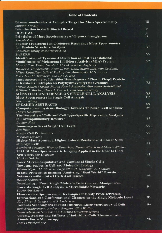
NOVAEDITORIAL Biomacromolecules: A complex target for mass spectrometrySimone Königiii-ivIntroduction to the Editorial Boardv-ix

REVIEWS

Principles of mass spectrometry of glycosaminoglycansJoseph Zaiapp 3-36Abstract:Structural variability resulting from protein post-translational glycosylation vastly increases the range of biological functions produced from the genome. The chemistry of glycans differs from that of proteins and dictates the use of specialized mass spectrometric techniques for successful analysis. Moreover, the unique chemistry of each glycoconjugate class drives the need for tailored analytical techniques. Glycosaminoglycans (GAGs) are linear glycans that coat the surfaces of all animal cells, bound to syndecan and glypican proteoglycan core proteins. There, they play developmentally critical roles as co-receptors for many classes of growth factors and growth factor receptors. Animal cells grow in organized extracellular matrix environments consisting of proteins, glycoproteins and proteoglycans. GAGs serve to create and support immobilized growth factor gradients and play structural roles in these extracellular matrices. Although, due to their acidity, fragility and polydispersity, GAGs have traditionally proven difficult to analyze using mass spectrometry, effective new approaches have emerged over the past few years. This review summarizes structural biochemistry of GAGs and the uses of mass spectrometry in their analysis, with an emphasis on recent developments that have proven effective.Fourier transform ion cyclotron resonance mss spectrometry for protein structureChristian Ihling and Andrea Sinzpp 37-53Abstract:FTICR (Fourier transform ion cyclotron resonance) mass spectrometry offers several advantages for the analysis of biological samples, including excellent mass resolution, ultra-high mass accuracy, and a wide mass range. This paper gives a short overview of recent applications and technological developments of FTICRMS, which are relevant to the field of protein analysis. Tandem mass spectrometry, "bottom up" and "top down" approaches for protein analysis as well as methods for studying protein conformations are described and illustrated with examples.

PAPERS

Identification of tyrosine-O-sulfation as post-translational modification of melanoma inhibitory activity (MIA) proteinMarcel P. de Vries, Nicole C.C.J.M. Rullens-Ligtvoet, Wouter J. Hoeberichts, Alain J. van Gool, Mario J.M. van Zeeland, Milou Kouwijzer, Gijs F. Verheijden, Annemieke M.H. Boots, Peter D.E.M. Verhaert, and Ebo S. Bospp 57-73Abstract:Matrix assisted laser desorption ionization time-of-flight mass spectrometry (MALDI-TOF MS) and isoelectric focusing (IEF) of pure recombinant human MIA (melanoma inhibitory activity) protein, as expressed in CHO cells, indicated that part of the protein was post-translationally modified. Whereas standard positive ion mode MALDI analysis of intact MIA revealed an additional peak 80 Da higher than the theoretical mass, IEF showed three major protein bands which could all unequivocally be identified as MIA isoforms by peptide mass fingerprinting. Negative ionization MALDI mass spectrometry on a quadrupole TOF instrument indicated that the differences between these isoforms were due to one or two 80 Da modifications confined to tryptic fragment 59-76. The identity of the 80 Da post-translational modification (PTM) was determined to be a Tyr-O-sulfation through enzymatic removal of the 80 Da moiety from MIA by arylsulfatase treatment, and through the absence of the 80 Da modification(s) in MIA harvested from cells cultured in the presence of chlorate, which is known to inhibit Tyr-O-sulfation. The PTM being sulfation was further corroborated by its metastability upon MALDI-TOF MS analysis. It is concluded that the CHO produced MIA is partially post-translationally modified by Tyr-O-sulfation at positions 69 and/or 70 (59LFWGGSVQGDYYGDLAAR76). Protein Tyr-O-sulfation, has been reported to serve various functions, including modulation of protein-protein interactions. Combination of this notion with the observation that the eukaryotic MIA protein shows only a weak binding towards laminin and fibronectin leads to the speculation that the putative in vivo metastasis promoting activity of MIA may be based on the interaction of MIA with an as yet unknown factor via Tyr-(O)-sulfation enhanced binding kinetics.Mass spectrometry identifies homologues of phasin Phap1 protein of Ralstonia Eutropha on polyhydroxybutyrate granulesMartin Zeller, Markus Pötter, Frank Reinecke, Alexander Steinbüchel, William I. Burkitt, Peter J. Derrick, and Simone Königpp 75-84Abstract:Many prokaryotes use polyhydroxyalkanoates (PHA) as storage compounds for carbon and/or energy in the form of water-insoluble inclusion PHA granules in the cytoplasm. The surface of such granules is covered by proteins which also affect their form and structure. Here, phasin PhaP1 protein was identified in high abundance on the grana surface of R. eutropha employing gel electrophoretic separation and mass spectrometry. Homologous phasins PhaP3 and Pha4 were visualized using 2D-PAGE in the PhaP1 knock-out mutant, but no evidence for the presence of PhaP2 could be found. All detected phasins exhibit isoforms whose structural differences are still a matter of investigation, although C-terminally truncated isoforms of PhaP1 were detected with FTICR-MS.
PROCEEDINGS OF THE 1st MÜNSTER CONFERENCE ON SINGLE CELL ANALYSIS 2004

INTRODUCTIONMass spectrometry in single cell analysisSimone Königp 87SPEAKER ABSTRACTSpp 89-97Computational systems biology: Towards ‘In silico’ cell models?Hergo HolzhütterThe necessity of cell- and cell type-specific expression analyses in cardiopulmonary researchLudger FinkImmunogenetics at single cell levelJan BuerSingle cell proteomicsNorman DovichiHigher mass accuracy, higher lateral resolution: A closer view of single cellsBernhard Spengler, Werner Bouschen, Dieter Kirsch and Martin KöstlerMALDI mass spectrometric imaging applied in the race to find new cures for diseasesMarkus StoeckliLaser micromanipulation and capture of single cells – New approaches in cell and molecular biologyYilman Niyaz, M. Stich, B. Sigmüller, R. Gangnus, K. SchützeIn situ proteomics imaging: Analysing "real world" protein networks within intact cells and tissuesWalter SchubertNanobiology: From single molecule detection and manipulation towards single cell analysis in microfluidic networksDario AnselmettiFluorescence spectroscopic techniques to study protein/protein interactions and conformational changes on the single molecule levelJörg Fitter, I. Gregor and J. EnderleinTowards scanning (near field) infrared laser microscopy of cellsErik Bründermann, Andreas Bergner, Götz Wollny, Jean-Sebastien Samson and Martina Havenith-NewenVolume, surface and stiffness of individual cells measured with atomic force microscopyHans Oberleithner