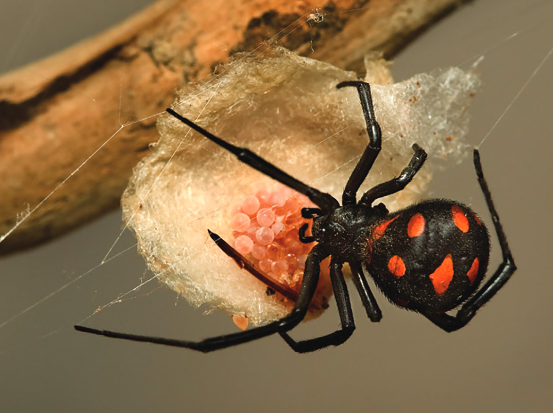
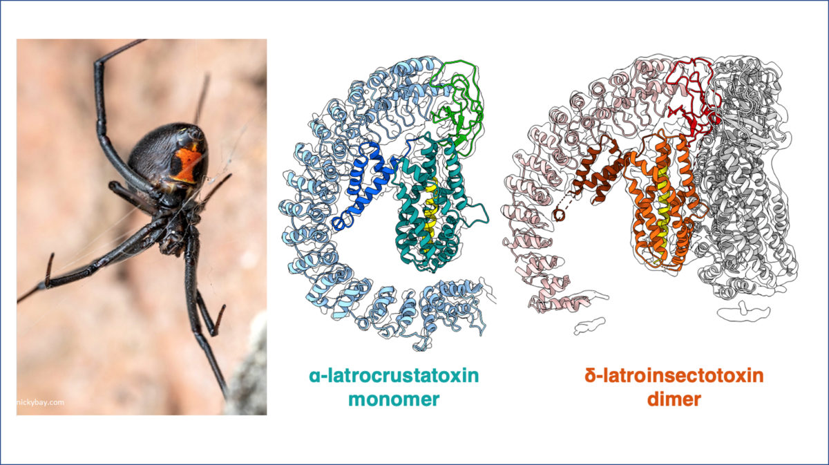
Black widows are the only spiders found in Europe whose venom is also dangerous to humans. The reason for this is the neurotoxin α-latrotoxin, which can trigger severe muscle spasms in humans by causing the uncontrolled release of neurotransmitters. To do this, a complex of the toxin forms a channel in the membrane of nerve cells.
The black widow is one of the most feared spider species. Its venom is a cocktail of seven different toxins that attack the nervous system. These so-called latrotoxins specifically paralyse insects and crustaceans, but one of them, the α-latrotoxin, targets vertebrates and is also poisonous to humans. It interferes with the signalling of the nervous system.
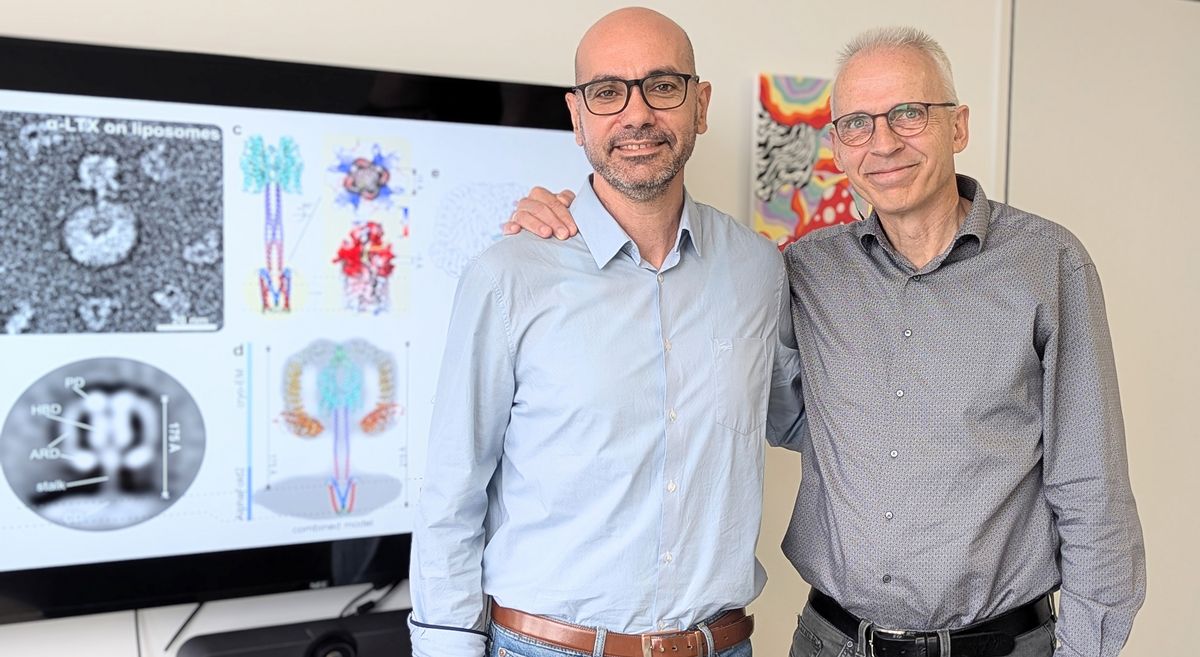
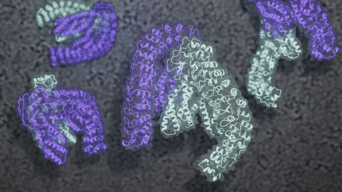
Every system has its waste disposal system. The cell organelles known as “peroxisomes” dispose toxic substances and fats in the human body, among other things, and, in doing so, they prevent serious illnesses. The “Pex” group of proteins (peroxisomes biogenesis factors) keep these “detox units” functioning properly – and a team of researchers at Münster University headed by Prof. Christos Gatsogiannis have now been the first to show, at the atomic level, how these highly complex processes proceed. The success story – now acclaimed with the study being published in the journalNature Communications– was made possible as a result of the University’s new high-tech microscope.
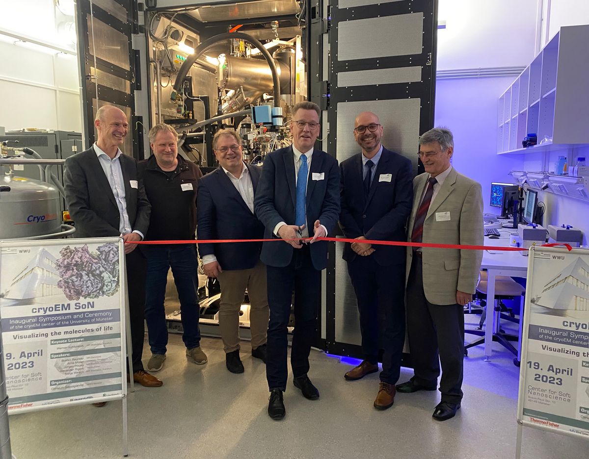
At an opening symposium with around 150 guests, a technically outstanding high-performance cryoelectron microscope ("cryo-EM") was officially inaugurated at the Center for Soft Nanoscience (SoN) of the Westfälische Wilhelms-Universität Münster (WWU) on 19 April (Wednesday). The device is one of the most powerful of its kind in the world and, under the direction of Prof. Dr. Christos Gatsogiannis, will in future be used by around 20 working groups and research associations from the disciplines of medicine, biology and chemistry. It is intended to contribute to the University of Münster maintaining and expanding its leading role in multiscale imaging internationally.
Many scientists have longed for it and now it is coming to be: following approval of their application to the German Research Foundation’s "Large-scale Research Equipment" funding programme, researchers from the University of Münster will receive equipment for high-performance cryogenic electron microscopy. The equipment will enable the researchers to make molecular processes visible – for example, in human cells – and to examine particles such as viruses and synthetic nanostructures three-dimensionally, down to their individual atoms. The German Research Foundation and the State of North Rhine-Westphalia are providing a total of 7.5 million euros for the preparatory equipment and the latest-generation high-resolution microscope, which will be located in a purpose-built laboratory at the Center for Soft Nanoscience (SoN).
When spider venom attacks the nerves: research team examines neurotoxin from a Black Widow
The team used cryo-electron microscopy to reveal the structures of toxins specific to insects and crustaceans
Photo: nickybay.com; Figure: Gatsogiannis Group
Although many people lose their nerve and panic when they see a spider, only very few of the creatures are actually dangerous. The Black Widow, however, is a force to be reckoned with: it catches its prey by means of nerve poison – to be precise, latrotoxins (LaTXs). Prof. Christos Gatsogiannis from the Institute of Medical Physics and Biophysics at Münster University investigated the substance – also with a view to medical applications. Using cryo-EM Gatsogiannis’ team succeeded in explaining the first structure of an LaTX. The research team’s findings have now been published in the Nature Communications journal. Here you can find the Publication in Nature Communications, the official press-release, also you are welcome to listen to a live interview with Prof. Dr. Gatsogiannis by “Radioeins” here.
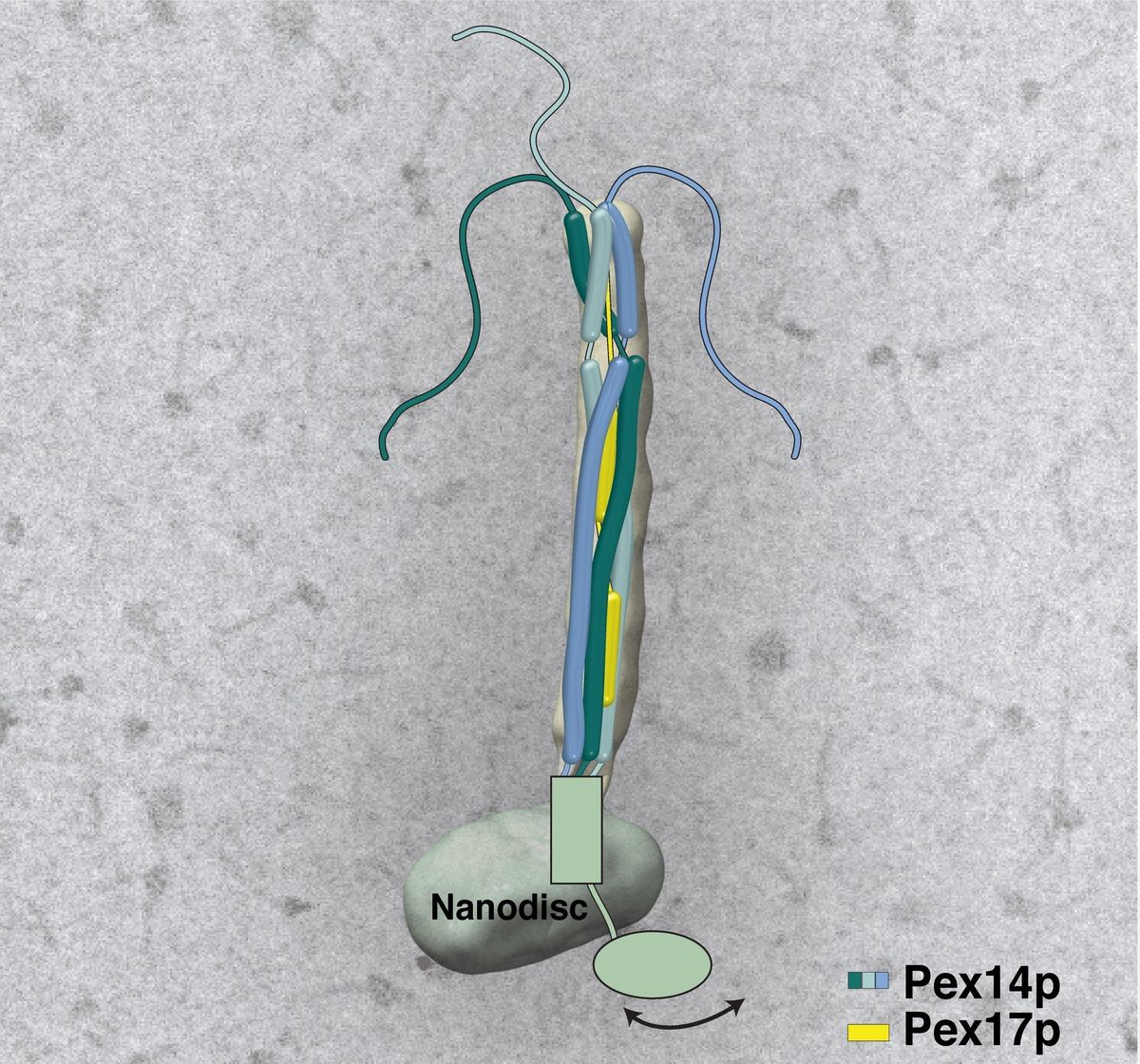
How peroxisomes "fish" for their enzymes: Scientists reveal the structure of the peroxisomal docking complex, December 2020
How peroxisomes "fish" for their enzymes: Scientists reveal the structure of the peroxisomal docking complex
Scheme of the peroxisomal Pex14pPex17p docking-complex, elucidated with electron cryo-microscopy (Fig.: Gatsogiannis)
Freiburg/Bochum/Münster - Peroxisomes are vital membrane-enclosed organelles that are found in every cell and are responsible, among other things, for its detoxification. For this purpose, they are equipped with an arsenal of enzymes. A team of scientists from the Universities of Freiburg, Bochum and Münster has now elucidated the first structure of the docking apparatus of peroxisomes, which captures enzymes for transport into the peroxisomes. The results were obtained in the DFG Research Unit "Structure and Function of the Peroxisomal Translocon" and have been published in the journal "Proceedings of the National Academy of Sciences of the United States of America" (PNAS).