8. Role of Ca2+ permeable channels in PDAC and PSCs
Objectives: The project aims to elucidate and to dissect, using tractable cell/organoid models in vitro, the mechanisms by which pH influences Ca2+ signals in tumour (PDAC) and stroma (PSC) cells as a function of the expression of genetic (KRAS2, SMAD4, TP53) drivers of PDAC.
In particular, we will focus on the remodeling of proteins forming store-operated Ca2+ channels (SOC) in “microenvironmental niche” associated with the induction of “apoptosis resistant” as well as “invasive” phenotype induced by the local microenvironment.
Migration, invasion and proliferation, apoptosis resistance, activation of PSCs and their secretions (e.g. growth factors, cytokines, matrix components) and cell-cell communication will be quantified.
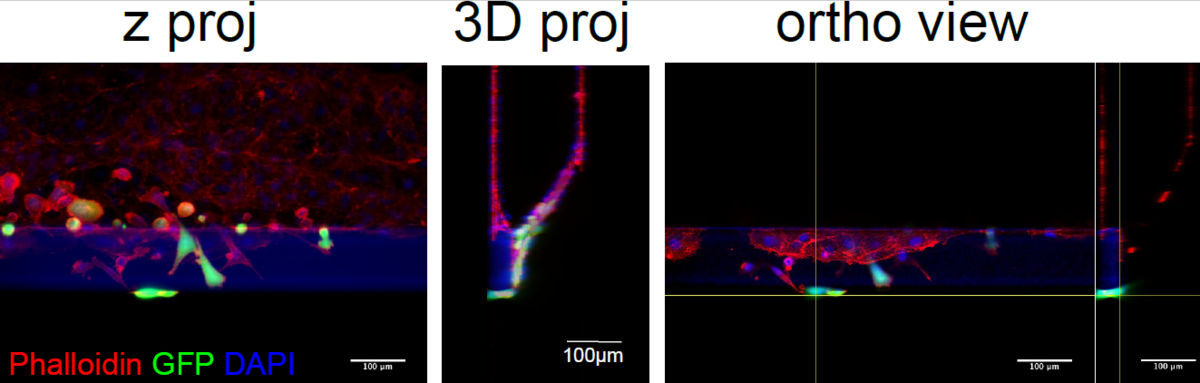
Figure 1: 3D microfluidic platform for perfusable vessel on-chip extravasation. Cancer cells were introduced in the perfused vessel and allowed to transmigrate for 12 hours. Figures represent different representations of confocal images. Left panel: z projection obtained averaging the intensity of the XYZ stack to generate a 2D projection. Central panel: 3-D reconstruction of the cancer GFP-stained cancer cells extravasation. Right panel: x, y and y, z orthogonal views of the perfused vessel containing cancer cells. Vessel were fixed and stained for Phalloidin (to visualize f-actin), DAPI (to visualize nuclei).