
News from the Institute of Medical Biochemistry (IMB)
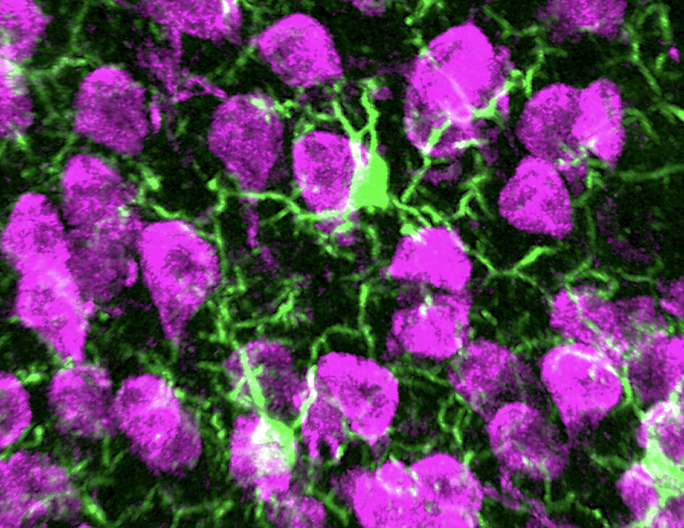
Our study in EMBO Reports identified an important molecular mechanism that helps microglia, important immune cells in the brain, develop and maintain healthy function in the central nervous system. We showed that the Arp2/3 complex, a protein complex that controls the structure and movement of cells by organizing actin filaments. We found that removing a key Arp2/3 component disrupted the normal maturation of microglia, preventing them from developing their characteristic branching shape and reducing their ability to monitor brain tissue effectively. Our findings show that actin remodeling plays a central role in keeping microglia flexible and balanced, while also helping prevent harmful or excessive immune activation in the brain.
Publication:
Safaiyan S, Frosch M, Bickel T, Monaco G, Dvir R, Madry C, Bosch LFP, Kierdorf K, Innocenti M, Priller J, Prinz M, Lämmermann T.
The Arp2/3 complex controls the development of homeostatic microglia. EMBO Rep 2026; 27(7):1696-1719.
Article Link: https://www.doi.org/10.1038/s44319-026-00721-8
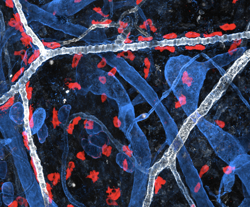
We discovered how specialized support cells in the skin help maintain healthy populations of mast cells, important immune cells involved in wound healing, allergy responses, and protection against infection. Our study identified dermal fibroblasts and cells surrounding blood vessels as the main sources of KIT ligand, a signaling protein essential for mast cell survival. We also showed that keratinocytes, cells at the upper surface of the skin (epidermis), are not critical for mast cell maintenance under normal conditions. The findings provide new insight into how different skin cell types create supportive local environments for immune cells and may help guide future research into inflammatory skin diseases and allergic disorders.
Publication:
Martzloff P, Testroet F, Salatino A, Ahmed A, Manke T, Biggs LC, Rognoni E, Bajenoff M, Mihlan M, Lämmermann T.
Mast cell homeostasis depends on KIT ligand from dermal fibroblasts and perivascular cells in mouse skin.
J Invest Dermatol 2026; Apr 13: S0022-202X(26)01023-7.
Article link: https://doi.org/10.1016/j.jid.2026.03.029
August 14th, 2024:
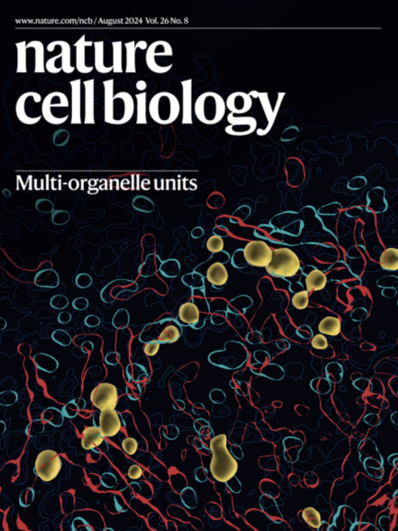
And congratulations again to Angelika and team for the cover of Nature Cell Biology (Volume 26 Issue 8, August 2024) highlighting their study on multi-organelle units.
Link: https://www.nature.com/ncb/volumes/26/issues/8

August 2nd, 2024:
New study on Mast Cell Intracellular Traps in “Cell”.
Happy to highlight our new study in Cell, which was led by first author Dr Michael Mihlan and senior authors Dr Angelika Rambold and Prof Tim Lämmermann. We discovered that mast cells can trap living neutrophils, forming an unknown cell-in-cell structure, before mast cells start using neutrophil components. Our study shows an unexpected functional liaison between these two cell types during allergic reactions. With great help from Manuel from IMB, several members of the MPI Freiburg team and numerous national and international collaborators.
Link: https://www.cell.com/cell/fulltext/S0092-8674(24)00774-8
Link: https://www.uni-muenster.de/news/view.php?cmdid=14211&lang=en
July 17th, 2024:
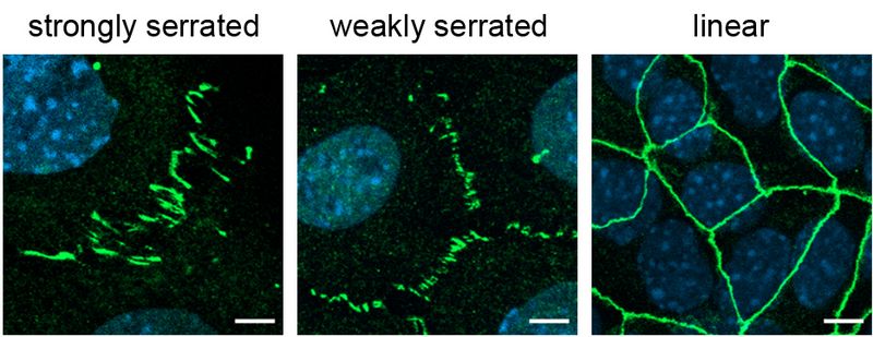
A new study by the team of Prof. Klaus Ebnet published in “Frontiers in Cell and Developmental Biology” has characterized the role of Rho GDP dissociation inhibitor 1 (RhoGDI1) in cell-cell contact formation of epithelial cells. The team found that RhoGDI1 regulates several processes associated with junction formation and function, including the development of cell-cell contacts, the formation of functional tight junctions, and collective cell migration. The study also identified the cell adhesion receptor JAM-A as a potential regulator of RhoGDI1 recuitment to cell-cell contacts. The study provides new insights into the molecular regulation of epithelial cell-cell contact formation and further underlines the role of JAM-A as a regulator of epithelial cell-cell junctions.

July 5th, 2024:
Visualizing the complex inner lives of macrophages.
Outstanding new study in Nature Cell Biology from the team of Dr Angelika Rambold, who will join our institute as independent group in early 2025. With “OrgaPlexing”, her team developed a novel multiplex imaging method that simultaneously illuminates several key organelle systems (mitochondria, endoplasmic reticulum, Golgi, peroxisomes, lipid bodies, lysosomes) in primary immune cells. Using this method to comprehensively map intracellular compartments, Angelika’s group identifies functional multiorganelle units (MOUs) that control intracellular cell metabolism and the production of inflammatory molecules in macrophages.
With great help from Frauke, Klaus and Manuel from IMB.
Link: https://www.nature.com/articles/s41556-024-01457-0