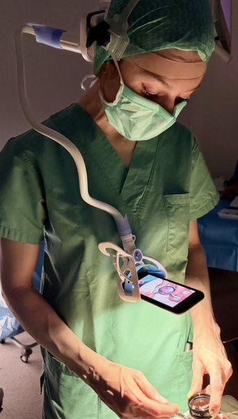
Study on Retinopathy of prematurity (ROP)
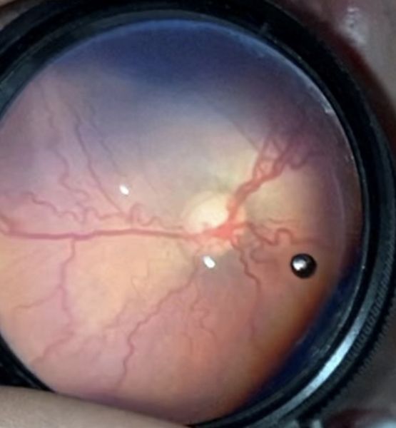
Under the supervision of Nataliia Mala, a feasibility study on wide-angle fundus imaging as part of screening for retinopathy of prematurity (ROP) is being conducted at our research centre. This is a prospective, single-centre, exploratory feasibility study without randomisation, without blinding, without a placebo and without any competing interventions.

Retinopathy of prematurity is a vasoproliferative retinal disease affecting immature preterm infants and is one of the leading causes of childhood blindness worldwide. Reliable documentation of the retina is therefore of great importance for diagnosis and monitoring of the condition’s progression. Binocular indirect ophthalmoscopy (BIO) is considered the gold standard for diagnosis. However, none of the systems currently available combines all the desirable criteria, in particular low acquisition costs, widespread availability, minimal discomfort for the child and high-quality wide-angle fundus documentation.

The aim of this study is to investigate whether a diagnostically useful wide-angle fundus collage of the retina in preterm infants can be reconstructed using a smartphone-based fundus video recorded during a standard BIO examination. In addition to technical feasibility, the study will examine, in particular, the retinal area captured, the image quality, and the diagnostic concordance of the findings with a reference BIO examination.

A total of 40 participants are to be recruited for the study, including 20 preterm infants and 20 healthy adult volunteers. The control group (adults) is intended for the technical validation of the algorithm and not as a diagnostic control group in the strict sense.

Participants must meet the following inclusion and exclusion criteria:

Group A (premature infants)

Inclusion criteria:

1. Preterm infants for whom ROP screening is indicated in accordance with the DOG/BVA S2k guideline 024-010

2. Sufficiently haemodynamically and respiratorily stable for a routine ROP screening examination at the scheduled time (to be determined by the attending neonatologist)

3. Written informed consent from the legal guardians.

Exclusion criteria:

1. Acute cardiorespiratory instability preventing a standard ROP examination at the scheduled time.

2. Pre-existing relevant ocular conditions unrelated to ROP that prevent fundus photography (e.g. pronounced tunica vasculosa lentis, corneal opacity, congenital cataract).

3. Lack of consent from the legal guardians for the processing of the video recording.
 

Group B (Adults)

Inclusion criteria:

1. Age ≥ 18 years.

2. Clear refractive media, adequate pupil diameter during mydriasis

3. Written informed consent.


Exclusion criteria:

1. Contraindication to mydriasis (e.g. known narrow anterior chamber angle, angle-closure glaucoma).

2. Pregnancy or breastfeeding.

3. Significant opacities in the ocular media, oil or gas tamponade, retinal detachment affecting more than one quadrant, or increased light sensitivity that impedes retinal examination.

4. Lack of consent.

Studienablauf

The examination essentially comprises the following steps:

  • Examination of preterm infants: Preterm infants are examined as part of the routine ROP screening programme. Following the standard pupil dilation and preparation, the standard BIO is performed. Immediately afterwards, a short video of the retina is recorded for each eye using a smartphone connected to the examination equipment via a special mount.
  • Examination of adult volunteers: Following pupil dilation, a standard examination of the retina is carried out. A short video of the retina is then recorded.
  • Analysis of the video recordings: The recorded videos are stored in pseudonymised form on a secure study server and subsequently analysed using specially developed software. First, suitable fundus images are filtered from the videos and assessed for image quality. Similar images are then merged and optimised.
  • Creation of a wide-angle fundus collage: Finally, the selected individual images are assembled into a coherent fundus collage based on the characteristic vascular patterns of the retina. The aim is to produce as comprehensive a representation of the retina as possible from the smartphone video recorded.

Further information can be found here on the DRKS website.

If you have any questions about the project, please feel free to contact the study management team.

Nataliia Mala
Klinik für Augenheilkunde
Universitätsklinikum Münster
T +49 15257910937
nataliia.mala@ukmuenster.de