Serend-IP, a spin-off from Münster University, offers lab services in nanoscale quantiftation of cell surfaces using AFM (Atomic Force Microscopy). The focus is on physiological barriers in vitro based on epithelial and endothelial models. Physiological relevance is achieved by high quality primary cells, isolated in cooperation with renowned research groups, Key competence of Serend-ip is the mapping of biological AFM-images through proprietary computer vision algorithms (nAnostic™-method - patented under US 8,798,935 / EP 2 435 829). The label-free quantitation of surface structures is operator-independent, robust and extremely accurate - down to a level of attoliters (10-18l). Various proof-of-concept studies were performed in contexts as different as toxicology, cardiovascular diseases, immunology, neurology, tumor and skin diseases.
Flanking assaying methods like cell migration, cell-toxicity, immunofluorescence etc. are conducted in close collaboration with scientific partners from universities and clinics across Europe to assure highest validity of the results. Projects are partially funded by (inter-)national grants (BMBF/EU). The company was founded in 2009, arising from research on cell mechanics of inflammation in endothelia. The first product on the market is the DERMATACT assay for skin health. The team is comprised of Dr. Christoph Riethmüller (scientific head), Dr. Armin Kramer (BioAFM Scientist), Jonas Franz (Medical Application) and Svetlana Izmailova, M.A. (Administration).
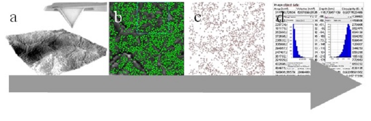
Figure 1: nAnostic-procedure a) the nanoscale topography of the cell sample is recorded via atomic force microscopy (AFM). b) Computer vision segments relevant objects (green) from background, c) Object map for morphometry and d) detailed number values are extracted, here only object count and local volume sum are displayed as histograms. For diagnosis, 10 arbitrarily chosen areas per sample are analysed and morphometrical data are averaged.
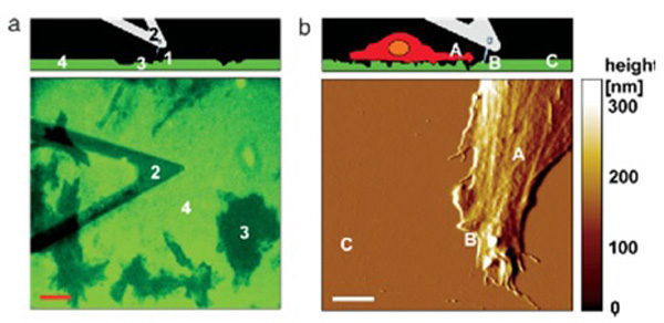
Figure 2: Morphometric quantitation of ECM degradation. a) the nanoscale topography of extracellular matix (ECM, green) as imaged via atomic force microscopy (AFM). A sketch (upper) demonstrates the imaging of a fluorescently labelled ECM (fluorescence image, lower). The degradation of the fluorescently labelled ECM is recognised as dark areas b) AFM-Imaging of a tumor cell on an a degradable extracellular Matrix (original image (lower panel) and schematic representation (upper). The total volume (AFM) of degraded ECM can be exactly calculated by nAnostic-Algorithms. Pictures taken from: Kusick et al. Jornal of Cellular Physiology 204: 767-774 (2005)