
Molecular imaging covers a broad spectrum of applications ranging from imaging of single cells or even subcellular structures in vitro to clinical diagnostic imaging in patients in vivo. Our aim is the non-invasive phenotypization of wild type, surgical and transgenic animal models of human disease using highly sensitive and high resolution dedicated small animal imaging technology. This core group offers both technology access and scientific expertise.
Positron emission tomography is a highly sensitive, quantitative imaging modality being capable of assessing molecular dynamics in vivo with nano-/picomolar sensitivity. The PET methodology available within the core unit is a dedicated high resolution small animal PET scanner with sub-milliliter resolution and a large field of view (28 cm * 16 cm). This camera is one of only four installations worldwide (quadHIDAC, Oxford Positrons Ltd., Oxford, UK). Due to cooperations with the Department of Nuclear Medicine and the Collaborative Research Center SFB 656 MoBil “Molecular Cardiovascular Imaging” at the University of Münster, this core unit is able to offer a wide range of different molecular imaging probes ranging from whole-body measurement of perfusion and metabolism down to cell imaging and imaging of targets being involved in oncological, inflammatory, neurological, cardiovascular and other diseases.Small Animal Computed Tomography (CT)
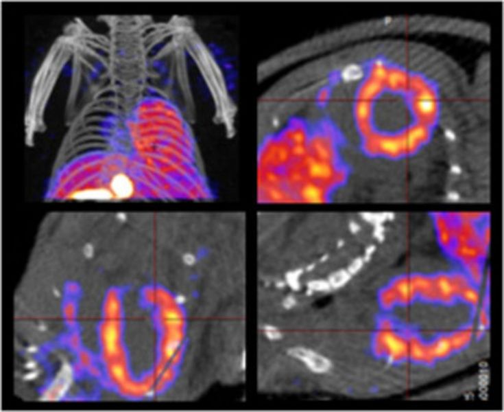
Computed tomography is an anatomic imaging modality with a very high spatial resolution. In the core unit group, this method is used primarily to provide anatomical information in correlation to the distribution of specific molecular imaging probes in PET and SPECT. In general, small animal CT alone offers a spectrum of applications similar to CT in the clinical setting.Integrated Small Animal PET/MRI
The core unit is also equipped with a 1-Tesla nanoScan PET/MRI, which is the first integrated in-line preclinical whole-body PET/MRI with a spatial resolution of 700 µm for PET and 100 µm for MRI that allows a simplified and optimized workflow for hybrid imaging. The anatomical correlation which is provided with this integrated PET/MRI improves localization especially in tumor or brain imaging studies. More sophisticated MRI sequences are possible in the dedicated small animal MRI system (imaging expert: Cornelius Faber), operating at a magnetic field strength of 9.4 tesla.
Small Animal Single Photon Emission Tomography (SPECT)
A dedicated multi-pinhole SPECT/CT system for animals having a sub-millimeter resolution and good sensitivity is installed in the core unit group (nanoScan SPECT/CT). The SPECT methodology is basically a gamma camera system which provides tomographic images. Since the gamma camera technology is the most common molecular imaging tool in patients a diversity of tracers/tracer kits are clinically available which can be directly used in small animal SPECT. This will substantially broaden the spectrum of molecular targets and methodology. Ex-vivo Bio-Distribution
Beside in vivo studies, questions concerning biodistribution and metabolism can be answered by ex vivo autoradiography, tissue counting studies.
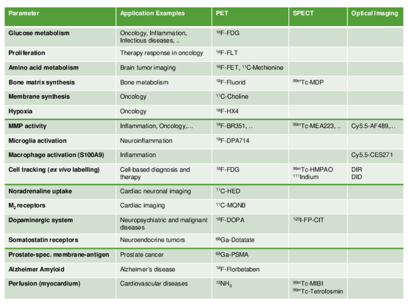
We offer a number of routine imaging protocols for cardiac, developmental or oncological models. An overview of available tracers and application examples is shown in the table on the left, also available as PDF. We are always open to broaden the spectrum of tracers established in our labs. If you are interested in imaging compounds not listed here or in evaluation of novel tracers please contact us.