The cells we are studying are primordial germ cells (PGCs), the founder cells of gametes in sexually reproducing organisms. Our group investigates the molecular and cellular mechanisms underlying germ cell fate establishment and maintenance, cell motility, directed cell migration and early stages of gonad development in the context of the live vertebrate embryo. Together, these processes ensure the differentiation of primordial germ cells into sperm and egg at the correct place in the embryo.
Germ cells thus serve as an excellent general model for studying medically relevant issues regarding the molecular mechanisms controlling cell fate decisions (relevant for gamete formation and oncogenic transformation), invasive cell migration (relevant for tumour cell metastasis and inflammation) and organogenesis (relevant for gonad function). Our primary model organism is zebrafish, where studying these processes from the earliest stages of development is facilitated by the extra-uterine development of the translucent embryo.
A developing zebrafish embryo (nuclei labeled in red) with germ cells (blue) migrating towards the sites where the gonad develops.
Developing zebrafish embryos (nuclei labeled in blue) with germ cells (red) migrating in the absence, or presence of the chemokine Cxcl12.
Molecular mechanisms controlling germ cell specification and fate maintenance
Our group identified proteins whose function is essential for cell fate maintenance and cell behavior. An example for such a protein is Dead end, a vertebrate-specific protein whose functions is associated with an increased rate of testicular tumours. We study the role of those proteins using genetic, optogenetic and genomic techniques, making use of different microscopy modalities in live zebrafish embryos.
Following the subcellular localisation of proteins and RNA molecules within phase-separated organelles in live embryos provides important information concerning the regulation over their role and function in germ-cell fate specification and maintenance.
Cell motility and chemokine-guided
migration
Similar to many other cell types in vertebrate
embryos, the germ cells polarize and migrate
directionally towards domains in the embryo where
the guidance cue Cxcl12 is expressed.
Exploring the mechanisms responsible for cell
motility and directed cell migration, we follow
the process of front-rear polarity establishment
using time-lapse movies. Employing a range of
genetic, reverse genetics and optogenetic tools,
we established hierarchical relationships among
molecules and activities in the cell that function
in transforming apolar non-motile cells into
polarized migrating cells. To obtain quantitative
description of a broad range of cellular features
relevant for cell motility we employ
machine-learning approach.
The molecular and cellular mechanisms controlling early gonad development
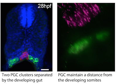
The precise positioning of organ progenitor cells constitutes an essential, yet poorly understood step during organogenesis. We use the germ cells and early gonad formation as a model for this important process. By the end of the first day of development, the PGCs reach their target and have to interact with somatic cells participating in gonad formation. Using live-cell imaging we found that the germ cells maintain their motility following their arrival at the gonad, yet remain in the region where the gonad develops. In addition to understanding the process of proper organ formation, determining the mechanisms confining motile cells to a specific site is highly relevant for understanding the process of cancer cell metastasis.
Analysis of cell behavior, analysis of the process in manipulated embryos and mathematical modeling revealed a role for tissues neighboring the gut in maintaining the bilateral arrangement of the cell clusters, cell cluster size and positioning.