News

JAM-A and cell cannibalism

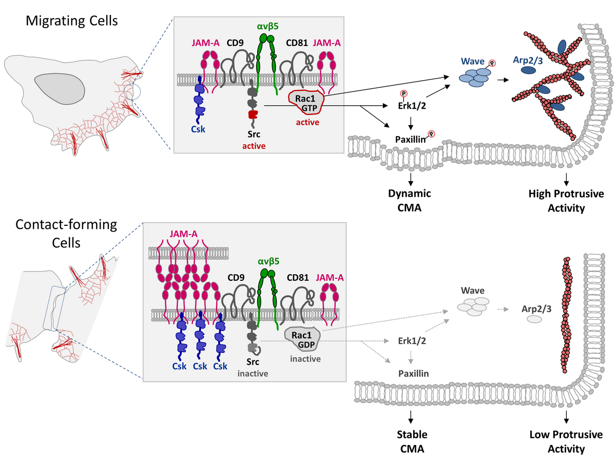
In our recent study we describe in more detail an observation that we made during an earlier study. In that study we had found that JAM-A limits cell motility and triggers a process called “contact inhibition of locomotion” (CIL) when migrating interact with other cells. This process is required to prevent cellular overgrowth and to ensure monolayer formation (see “News” section below: “JAM-A makes contact inhibition”).

When we analyzed cells migrating on micropatterns we observed that JAM-A-depleted cells not only migrated across other cells but formed cell-in-cell structures, as if cells would feed on each other through cell cannibalism. Cell-in-cell structures between non-phagocytic cells are frequently observed, and various types of cell-in-cell structures are described in the literature.

We characterized the type of cell cannibalism observed after JAM-A depletion as entosis. During entosis, tumor cells internalize other tumor cells to get access to the energy resources of the internalized cell. The onset of entosis after JAM-A depletion strongly correlated with a loss of CIL and involves the same molecular mechanism.

We found that the suppression of entosis by JAM-A correlates with the ability of JAM-A to regulate CIL, and that it involves the same molecular mechanisms. Our observation, thus, indicate that JAM-A not only regulates inhibition of cell motility in response to cell-cell contact formation but that a loss of CIL is linked to a process of cell cannibalism called entosis. This study is published in iScience.
 

Two MCF7 tumor cells – one JAM-A KD cell (green) and one wildtype cell (red) –  in a fierce battle. Note that the red cell repeatedly escapes from the green cell’s clinch. In the majority of cases, the JAM-A KD cells ended up as the winner and degraded the wildtype cell within a vacuole.  


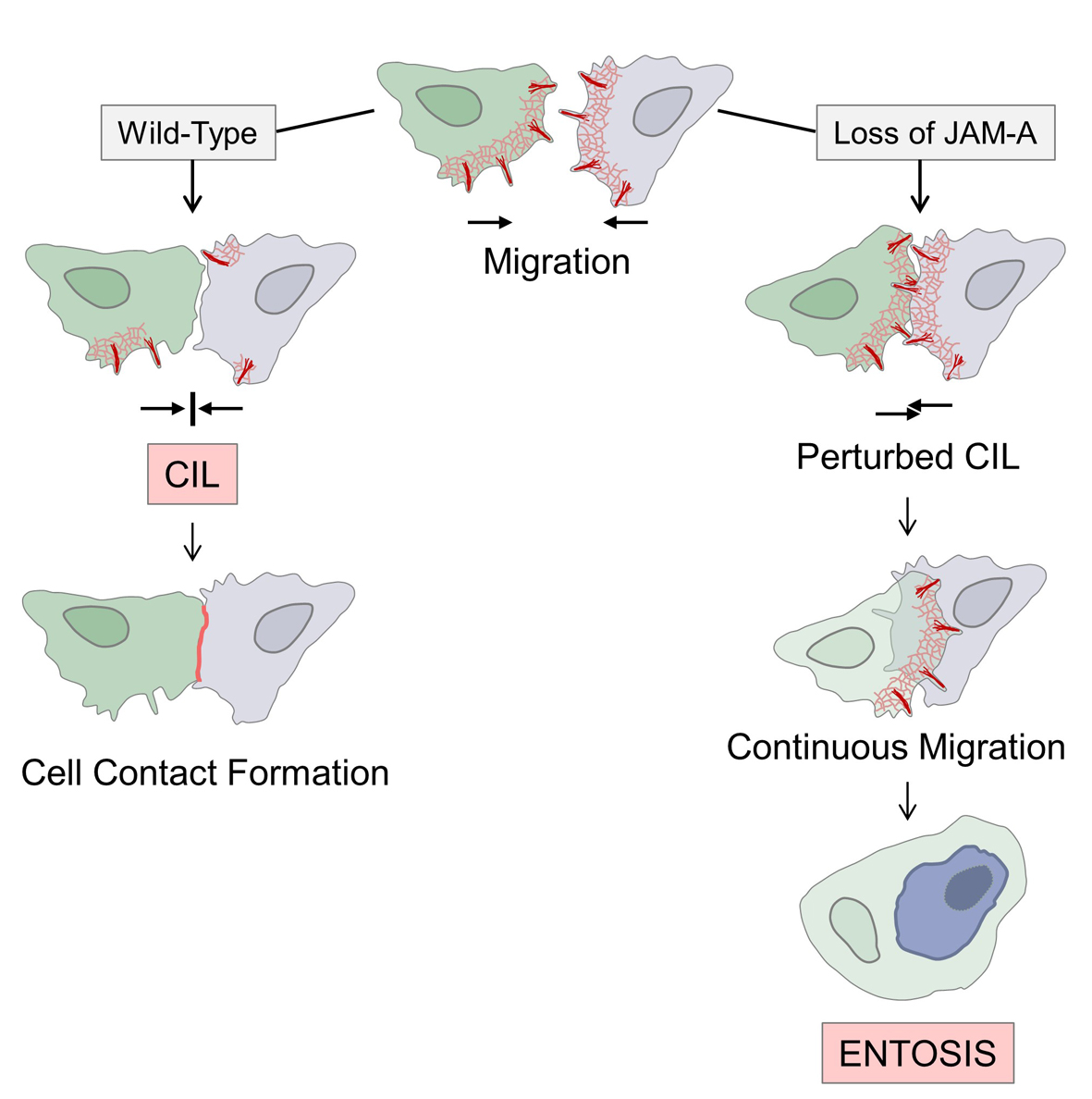
Left (wildtype condition): Contact-forming migrating cells downregulate protrusive activities (normal CIL response) and form cell-cell contacts. Right (JAM-A knockdown condition): Contact-forming cells fail to downregulate protrusive activities (no CIL response) and continue migration. In many cases, one of the two cells cell is engulfed by the other cell through entosis. 


Cell adhesion receptor TMIGD1 brings up the brush border

We describe an unexpected function of an adhesion receptor with a high structural similarity to Junctional Adhesion Molecules (JAMs). To our surprise, this adhesion receptor – called “Transmembrane and immunoglobulin domain-containing protein 1” (TMIGD1) is localized at the brush border and helps to connect individual microvilli with each other.

Intestinal epithelial cells contain a densely packed array of microvilli at their apical membrane domain which together form the brush border. Microvilli amplify the surface of the small intestine by a factor of 10 to 15, which allows efficient nutrient absorption. Microvilli are remarkably uniform in length and are very regularly packed. This optimal arrangement requires adhesion between individual microvilli, which is partially mediated by a protocadherin-based adhesive complex at their tips, the intermicrovillar adhesion complex (IMAC).

In our study, we identify a second adhesive complex at microvilli, which we call IMAC2. This complex is based on TMIGD1 and localizes to the subapical region of microvilli. TMIGD1 is recruited to microvilli by two scaffolding proteins, NHERF1 and NHERF2. Through its interaction with NHERF1, TMIGD1 is linked to ezrin which in turn links the entire complex to the underlying actin cytoskeleton. We find that deletion of the Tmigd1 gene in mice results results in a strongly perturbed brush border, accompagnied by a loss of intermicrovillar adhesion and microvillar blebbing. Our observations also suggest a molecular mechanism that could explain why the highly dynamic growth and shrinkage of microvilli (click on the Inset for visualization) is restricted to the apical region of microvilli. Thanks to Christian and Eva for this great work, and thanks to Lilo for the beautiful EM pictures. The study is published in Science Signaling. Here is the link to the reprint:  https://www.science.org/stoken/author-tokens/ST-729/full

Adhesion complexes at microvilli. MV tip: Microvilli are linked at their tips by an intermicrovillar adhesion complex (IMAC). The adhesion is mediated by trans-heterophilic interactions between two protocadherins called “Cadherin-related family member 2” (CDHR2) and CDHR5, which are linked to the underlying actin cytoskeleton through the scaffolding proteins USH1 and ANKS4B, and the unconventional myosin MYO7b. A second intermicrovillar adhesion complex  (IMAC2) is localized at the proximal base region of microvilli. The IMAC2 is based on trans-homophilic interactions between TMIGD1 molecules on adjacent microvilli. TMIGD1 molecules interact with the cytoplasmic scaffolding proteins E3KARP (NHERF2) or EBP50 (NHERF1). The interaction of TMIGD1 with EBP50 is triggered by active Ezrin, which links the complex to the actin cytoskeleton. Microvilli are very dynamic structures (see video below for animation). Our findings strongly suggest that the IMAC2 regulates microvilli dynamics. 


Dynamics of microvilli formation. Microvilli are dynamic structures. In cultured epithelial cells, microvilli undergo phases of growth and shrinkage with an average life cycle of 12.1 +/- 5.6 min. Thanks to Lynn Glaesner-Ebnet for the animation. 


JAM-A makes contact inhibition

We uncover a mechanism through which cells downregulate their motility when they collide with other cells.

Migrating cells display vivid mobility, which is characterized by a high protrusive activity and by dynamic cell-matrix interactions. When migrating cells encounter other cells, they typically stop their protrusive activities at these site and stabilize their interaction with the extracellular matrix at cell-matrix adhesion, which results in a downregulation of their motility. This process is commonly referred to as “contact inhibition of locomotion”. Contact inhibition of locomotion allows cells to become integrated into a tissue and prevents invasive growth.

In our study, we find that the cell adhesion molecule JAM-A exists in a multimolecular complex with tetraspanins CD9 and CD81, and with αvβ5 integrin. The two tetraspanins connect JAM-A with αvβ5 integrin, thus generating spatial proximity between JAM-A and αvβ5 integrin. This enables JAM-A-bound Csk to inhibit the activity of αvβ5-bound Src. We also find that JAM-A limits the activity of the Rho family GTPase Rac1, most likely in cooperation with CD81. JAM-A thus limits the activities of two central regulators of cell motility, Src and Rac1. As a consequence of elevated Src and Rac1 activities after JAM-A depletion, cells fail to stop migration when they collide with other cells but instead migrate across these cells, a behaviour that is typical for tumour cells. Our work thus provides a deeper insight in the regulation of contact inhibition of locomotion in tumour cells.

Migrating cells: JAM-A is localized at cellular protrusions and limits – but does not completely inhibit – the activities of Src and Rac1. Active Src and Rac1 act on Erk1/2, paxillin and the Wave complex to keep cell matrix interactions (CMA) dynamic and to promote protrusive activity. Contact-forming cells: JAM-A homophilic interactions result in JAM-A clustering at cell-cell contact sites. Enhanced recruitment of Csk by Tyr280-phosphorylated JAM-A leads to efficient inhibition of Src that is bound to αvβ5 integrin. Through its association with CD81, JAM-A also inhibits Rac1. Inhibition of Src and Rac1 results in inhibition of their downstream targets Erk1/2, paxillin, and the Wave complex. As a consequence, cells stabilize their interaction with the extracellular matrix at CMAs and downregulate their protrusive activity.