


Structural Metabolism of Inflammation
Metabolic programming emerged as a central mechanism in the regulation of adaptive and innate immune cell function. Intracellularly, this rewiring relies on the rapid adaptation of different organelle systems, specialized metabolic sub-compartments, which can arrange themselves in functionally interconnected networks to from metabolic circuits.
The Rambold lab focuses on untangling the identity and mechanisms driving this structural layer of metabolic control and how organelle and their networks shape immune cell functions during conditions of inflammation and infection on the single cell and populational level.
Goals
Immune cell defects leave us open to attacks from bacteria, viruses and more. However, an overacting systems is, in many cases, as harmful and dangerous as one that stops working. By deciphering how organelle-networks instruct immune cell metabolism and function, our work is aimed at identifying novel avenues that may help us combat immune-related diseases.
Approach
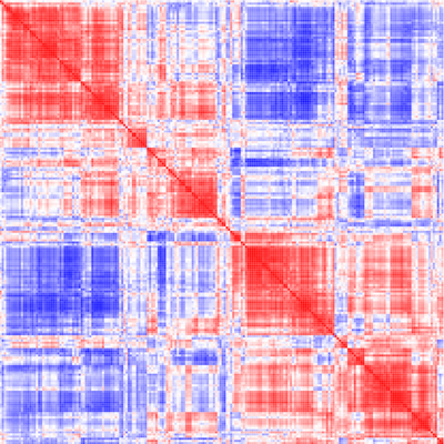
We follow an interdisciplinary research approach at the interface between immunology, cell biology and metabolism. We are combining cutting edge organelle and metabolic imaging approaches (advanced live and super-resolution imaging)
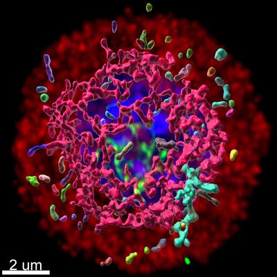
and analysis with metabolomics and RNA-sequencing techniques (both at bulk and single organelle/cell level) on primary immune cells for the identification of orga-metabolic networks. Candidate pathways are tested in relevant animal models, human disease models and/or patient material.
Projects
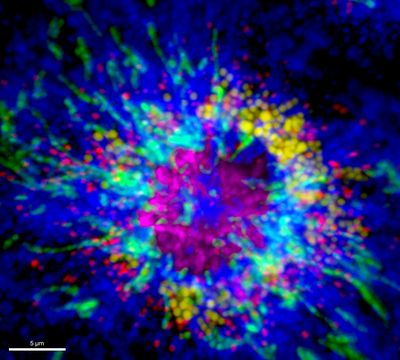
Immune cell function is instructed by specific rewiring of metabolic programs. Changes in the activity of metabolic pathways are key to sustain energy supply and generate building blocks for the synthesis of macromolecules. Beyond this, metabolites have also been shown to instruct a wide spectrum of cellular processes, including protein function, epigenetic state, pathogen defense and to serve as inter-cellular signaling cue, together illustrating the power of metabolic programming in shaping immune cell function.
In recent years, it has become clear that the majority of metabolic pathways is controlled by the function of different organelles. These discrete membrane-bound compartments are specialized metabolic machines and allow for the separation of often incompatible metabolic processes. For example, lipid metabolism is distributed between the endoplasmic reticulum and mitochondria for lipid synthesis, lipid droplets for storage, mitochondria and peroxisomes for β-oxidation, and lysosomes for lipid hydrolysis and recycling. To integrate the compartmentalized metabolic sub-processes and allow cells to reprogram their metabolism, the activities of these organelles must be coordinated.
Based on mostly pair-wise organelle studies from yeast and non-immune cells, it is becoming clear that many, potentially all, organelles are able to communicate with one another. However, we are still missing clear concepts on which organelles are used to form immune-relevant metabolic networks and how they shape immune cell function when cells adapt their function e.g. during conditions of inflammation and infection. In particular, altering the flow of nutrients and metabolites within a cell requires the coordinated interplay of several organelle systems, which can occur through two principle modes of communication: (1) direct physical contact through special membrane contact sites or organelle fusion to facilitate the transfer of metabolites, and (2) indirect communication between organelle systems through the activation of transcriptional programs or the secretion of bioactive signaling molecules to adapt intra- and inter-cellular organelle activity. Although it is meanwhile established that shifts between metabolic programs and levels of specific metabolites are essential for the regulation of immune cell activity, the functional aspect of interconnected organelle networks and their link to metabolic reprogramming in immune cells and immune disorders has largely been neglected.
In our research, we are particularly interested how organelle-driven metabolic programs/circuits control (a) the activity of individual immune cells under homeostatic conditions, (b) the sensing, detection and elimination of pathogens and (c) the communication within one or between several immune cell populations to coordinate their responses during inflammation and infection.
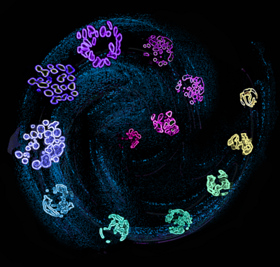
Mitochondria are key signaling nodes that can shape immune cell function by releasing ATP, ROS, mitochondrial DNA, TCA cycle intermediates, cytochrome C and more. Many of these functions are dependent on the dynamism of this organelle. Mitochondria continuously move inside cells and shape their morphology by undergoing controlled cycles of fission and fusion. In non-immune cells, we and others identified that mitochondria form hyperfused networks in response to limited nutrient availability (Rambold, PNAS 2011). This adaptive response protected mitochondria from autophagosomal degradation (Rambold, PNAS 2011), enabling them to support autophosomal biogenesis (Hailey, Rambold, Cell 2010). In addition, we found that fusion was essential to ensured proper intra-mitochondrial metabolite trafficking and respiration (Rambold, Dev Cell 2015).
These results prompted us to investigate if this mitochondrial hyperfusion is conserved in immune-cell transitions that undergo phases of ‘pseudostarvation’, e.g. the differentiation of effector to fatty acid oxidation reliant memory T cells. Morphological mapping studies revealed that effector T cells contained primarily fragmented mitochondria, whereas mitochondria in memory T cells were fused into networks. Inhibiting mitochondrial network-formation by depleting the fusion protein OPA1 resulted in cristae expansion and altered metabolic state. Most strikingly, we found that the failure to fuse mitochondria abolished the differentiation of memory T cells in vitro and in vivo (Buck, Cell 2016). As such, our data suggest mitochondrial remodeling as signaling mechanism that instructs T cell metabolic programming and function.
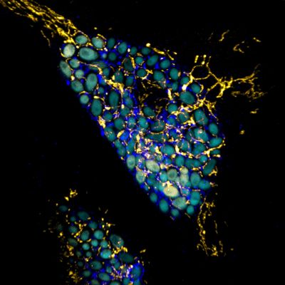
Phagolysosomes are primary organelle system to sense and kill many invading pathogens. Beyond this the lysosome has recently emerged as a dynamic and central metabolic hub. It recycles nutrients from digested material, can regulate metabolic signaling nodes (e.g. mTOR) and engage in extensive direct or indirect interactions with other membrane-bound organelles. This includes stable physical junctions with other membrane compartment, which likely provide direct routes for the exchange of lipids, ions and other metabolic signals.
Current work in the lab is focused on untangling how direct and indirect lysosomal communication controls macrophage functions during inflammation and infection. We are further investigating how defects in lysosomal functions contribute to immunological disorders. Of particular interest is hereby the lysosome-linked Chediak-Higashi syndrome, a primary immunodeficiency, whose patients suffer from recurring bacterial infections and a, causally unresolved, secondary hyper-inflammatory phase.
Based on mostly pair-wise organelle studies from yeast and non-immune cells, it is becoming clear that many, potentially all, organelles are able to communicate with one another via direct- or indirect interaction modes. In this project we aim at gaining a comprehensive understanding about large-scale organelle communication networks (via direct and indirect communication modes) and delineate how they are used to adapt organelle homeostasis and metabolism in healthy and diseased immune cells.
We are using targeted and systems approaches, combining high-resolution multicolor imaging with gene expression, proteomic and metabolic profiling on the population and single cell/organelle level to gain an integrated view of organelle-networks in primary macrophages, during inflammation, infection and resolution processes. By comparing our results with macrophage responses from models of human organelle-linked immune disorders, we aim at untangling primary defects from secondary spreading effect along the organelle networks. These may provide novel intervention points in the effort to ameliorate immune cell defects in affected patients.