General
Endocytosis
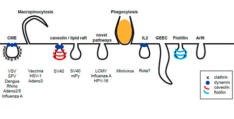
Endocytosis is an essential process of eukaryotic cells that is needed to internalize extracellular particles and solutes. Phagocytosis, the uptake of large particles, is typically restricted to specialized cells. In contrast, the uptake of small particles and solutes (pinocytosis) occurs virtually in all eukaryotic cells. For a long time it has been thought, that pinocytosis is mainly comprised of clathrin-mediated endocytosis. However, current research indicates a complex network of diverse ongoing and triggered pathways.
The pathways that viruses use for entry involve clathrin-coated vesicles, caveolae, macropinosomes, or further still poorly characterized pathways. Currently, we are particularly interested in a novel endocytic pathway used by Human Papillomaviruses type 16 (HPV-16) that bypass the classical organelles of established endocytic pathways.
Papillomaviruses
Research on the infectious potential of papillomaviruses evidently is important on its own account. Infection by certain papillomaviruses causes - besides the occurrence of warts in skin and mucosal tissues - fatal cervical and anogenital cancers. Despite screening efforts and the advent of vaccination strategies, cervical cancer is still the third most abundant cancer for women worldwide, and thus continues to be a major health threat, especially in developing countries.
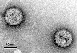
Papillomaviruses are small DNA viruses consisting of two structural proteins, L1 and L2, that make up the icosahedral (T=7) capsids. Little is known about the entry of the high risk papillomaviruses mainly due to the great difficulties growing infectious particles in tissue culture systems. For our studies, we use so-called pseudovirions that resemble infectious particles but lack the viral DNA. Instead, the particles harbour a reporter plasmid that is used to monitor efficient endocytic entry.
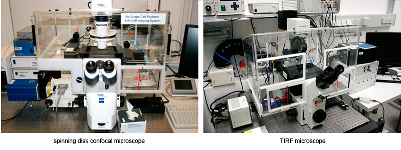
Equipment