
Methodological Developments in Functional MRI
Functional MRI is a diagnostic technique used in both clinical and preclinical research which provides high-contrast, noninvasive imaging. It visualizes the regional vascular response to local neuronal activity, which consists of changes in blood oxygenation and blood flow and lasts for several seconds. By detecting these vascular changes across the entire brain, conclusions can be drawn about regional neural activity and, subsequently, the activity of cerebral networks.
Our methodological developments are focused primarily on addressing two fundamental challenges in fMRI.
On the one hand, the temporal resolution in fMRI must be improved to visualize the interactions between different brain regions within identified networks. This is achieved by using the “line-scanning” method, in which the slow brain-wide imaging is replaced by targeted rapid scanning of regions of interest. Combined with the resulting ability to precisely identify the onset of the regional vascular response to neural activation (Cleppien 2022), this enables the visualization of the temporal course of interactions between different brain regions.
The second fundamental problem with fMRI that we address concerns the causal relationship between the measured signal of the vascular response and the underlying neural activity of interest. For example, the neural activity of interest may be masked by other neural activities, thereby influencing the vascular response. To allow for the distinct identification of the vascular response in the measured fMRI signal to the neural activity of interest, we use the bimodal approach of fast fMRI in preclinical diagnostics, combined with simultaneous optical fiber-based detection of local intracellular calcium concentration changes in a cellular region, since the calcium concentration changes measurably during neural activity. By integrating the neural activation identified in the calcium signal into the fMRI analysis process, only the vascular response to the neural signal of interest is specifically investigated (Cleppien 2022). This enables the visualization of the brain-wide signature of the local neural activity of interest using fMRI.
This bimodal diagnostic approach was applied to slow oscillations in the brain. This neural activity occurs during deep sleep and propagates from a starting point as a wave of neural activity across the cortex. Using this bimodal diagnostic method, it is now also possible to visualize the propagation of the vascular response to this neural activity (Cleppien 2025), as shown schematically in Figure 1.
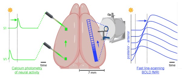
Figure 1: A sensory stimulus triggers a slow oscillation that travels across the cortex and can be detected using optical calcium imaging (green). The vascular response triggered by this distinct neural activity follows the neural activation and can be measured using fast “line scanning” fMRI (blue).
Our application of the bimodal approach to human diagnostics combines fMRI with EEG recordings, which can indeed measure neural activity but with significantly lower spatial resolution (Ilhan-Bayrakcı 2022). In the diagnosis of slow oscillations, this bimodal approach now allows for the visualization of different fMRI stimulation patterns in the human brain which depend on the various types of slow oscillations (Ilhan-Bayrakcı 2026). As neurophysiological markers, these stimulation patterns now enable conclusions to be drawn about the underlying integrity of neural networks.