
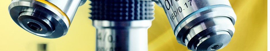
DHM - DIGITAL HOLOGRAPHIC MICROSCOPY
Label-free quantitative imaging of living cells and tissues
Technology & Features
Digital holographic microscopy (DHM) provides label-free quantitative phase imaging of biological specimens for modular integration into common optical microscopes. The reconstruction of digitally captured holograms is performed numerically. This feature allows multi-focus imaging of specimen parts in different layers and subsequent autofocussing from single captured digital holograms without mechanical focus realignment. DHM requires only low light intensities for object illumination and thus minimizes the interaction with the sample. This is an important advantage for minimally invasive quantitative long-term monitoring of living cell cultures. DHM phase contrast imaging simplifies automated object tracking and image segmentation for quantification of cell migration and morphology by extraction of absolute biophysical parameters such as cellular volume, thickness and dry mass. The evaluation of quantitative phase images also provides access to the cellular refractive index which is related to the cellular water content, intracellular solute concentrations and tissue density.
Biomedical Applications
DHM allows multimodal quantitative imaging of living cells and unstained tissue sections and can be utilized to investigate the influence of drugs, toxins and nanoparticles on cell morphology, growth and motility as well as for quantification of inflammation mediated tissue density changes. Moreover it enables the analysis of the cellular response to optical manipulation, the quantification of wound healing in-vitro and the observation of cancer cell migration in 3D tissue models.
Related article at www.photonics.com:
B. Kemper, "Digital holographic microscopy enhances cytometry and histology", Europhotonics Winter 2019, 20-23 (2019)
 BMTZ Brochure on Label-free Imaging