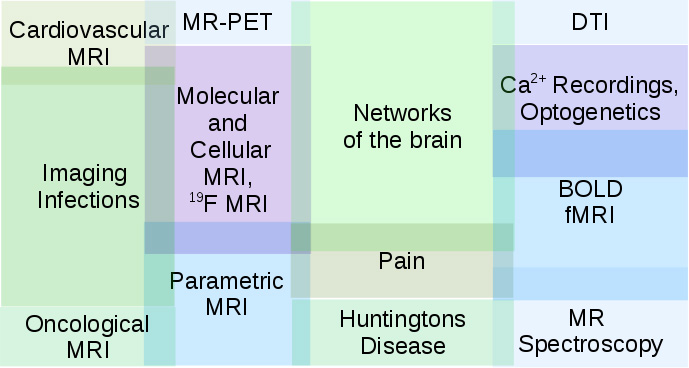
Magnetic Resonance Imaging (MRI) is one of the most important imaging modalities in medical diagnostics. MRI was first developed and implemented independently by P. Mansfield and P. Lauterbur in 1973. Today MRI is highly developed affording image resolutions on the order of 0.1 mm in human subjects and in the micrometer range in small objects, such as fixed specimens or single cells. In particular parallel acquisition strategies have reduced the minimum scan time and allow for acquisition of complete images in less than 30 msec, which makes real time visualisation of tissue movements feasible. For a large number of pathologies complete scan protocols are available for clinicians to use MRI in a black box manner for diagnosis of disease. We are working on the refinement of existing and develoment of novel MR methods to improve biomedical diagnostics and open new analytical options. The major modalities we employ are:
- Cellular / molecular MRI
- bacteria tracking
- SPIO-labelled macrophage / T cells
- 19F MRI
- Heteronuclear proton MRI
- Neuroimaging
- functional MRI
- BOLD fMRI
- BOLD fMRI + Ca2 recordings
- BOLD fMRI + optogenetics: ofMRI
- in vivo MR spectroscopy
- DTI
- functional MRI
- PET-MR
- parametric MRI: relaxation time mapping, vessel size imaging,...
- Neural networks in the brain
- structural and functional organization of neuronal networks
- pain processing
- huntingtons' disease
- Infection and inflammation imaging
- bacterial infections
- immune cell tracking


