STORM
(d)STORM:
(direct) Stochastic Optical Reconstruction Microscopy (dSTORM) is a new superresolving technique which enables resolution beyond the classical diffraction limit by localizing fluorescent single molecules with high precision. This requires special fluorescent dyes which switch between a fluorescent and a non-fluorescent state. During measurement, a small number of dye molecules are continuously activated in a stochastic manner, allowing precise position estimation. After photobleaching or switching back into the dark state, the next cycle of activation and detection follows. The combination of the gathered molecule positions results in a superresolved reconstruction of the structure of interest. Since this method requires longer acquisition times than classical fluorescence microscopy, the samples usually have to be fixed.
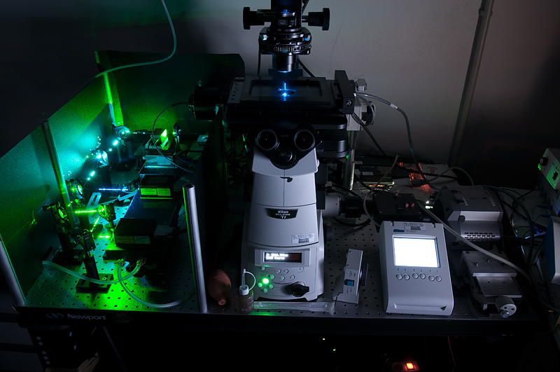
We have three different setups for (d)STORM microscopy at our disposal. The first one is a commercial Nikon Eclipse Ti-E TIRF microscope which combines the high lateral resolution of STORM with the vertical resolution of TIRF microscopy.
The second setup is a customized Nikon Eclipse Ti-E microscope which employs so-called astigmatic detection (B. Huang, W. Wang, M. Bates, and X. Zhuang, Three-dimensional super-resolution imaging by stochastic optical reconstruction microscopy. Science 319, 810 (2008)). Here, estimation of the axial coordinate is enabled by an additional cylindrical lens in the beam path which leads to a z-dependent ellipticity of single molecule images. This is used to estimate the axial coordinate with high precision, allowing 3D imaging.
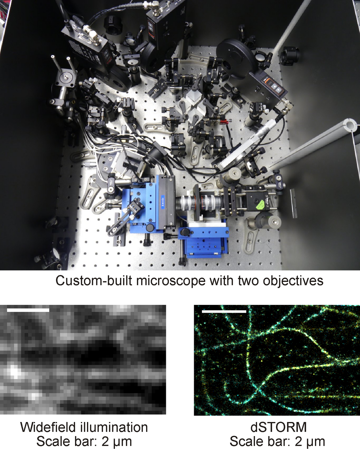
The third setup is an exceptional custom-built modular microscope with simultaneous detection via two objectives, offering even higher resolution. Here, two different options are implemented for 3D imaging, firstly astigmatic detection (K. Xu, H. P. Babcock, and X. Zhuang, Dual-objective STORM reveals three-dimensional filament organization in the actin cytoskeleton. Nat. Methods 9, 185 (2012)) and secondly interferometric detection (G. Shtengel et al. Interferometric fluorescent super-resolution microscopy resolves 3D cellular ultrastructure. Proc. Natl. Acad. Sci. U.S.A. 106, 3125 (2009), as well as D. Aquino et al. Two-color nanoscopy of three-dimensional volumes by 4pi detection of stochastically switched fluorophores. Nat. Methods 8, 353 (2011)). The latter method features a high axial resolution. It is however only suitable for thin and homogeneous samples. As a special feature, our microscope is equipped with glycerol-immersion objectives. They suppress aberrations. For details see: N. C. Schmidt, M. Kahms, J. Hüve, and J. Klingauf, Intrinsic refractive index matched 3D dSTORM with two objectives: Comparison of detection techniques. Sci. reports 8, 13343 (2018).