Serend-IP has more than a decade of experience in quantification of cell surface topography on the nanoscale. On top of (bio-)physical expertise on state-of-the art analytical techniques (AFM, nanomechnical mapping, electrical cell impedance spectroscopy, TIRF, FRET and 4Pi-fluorescence imaging etc.), a broad knowledge of physiologically relevant in-vitro testing methods is present (Cell migration, Invasion / Transmigration Assays, biochemical toxicity assays etc.). Special expertise has been gained on cell cultivation protocols for a large range of epithelial and endothelial models. The computer vision algorithms and neuronal networks necessary for the nAnostic-assays have been developed in house and it is the first and only available solution for biological AFM-images; this is the key competence of partner 2 (Serend-ip GmbH). Dr. Riethmüller has successfully participated in national (IMF) and coordinated international research cooperation projects (BMBF).
CEO Dr. Christoph Riethmueller has developed the nAnostic™ method for nanoscale phenotyping of cells. The start-up company Serend-ip GmbH is a spin-off from the institute of physiology at the University of Muenster which offers nAnostic-services to researchers in pharmacology and related disciplines. Since the proprietary pattern recognition software in early 2011 has reached a satisfying level of performance, the method is being validated in various biological contexts and cell models. This is performed in close collaboration with partners from universities and clinics in Germany and Western Europe. Serend-ip offers highly specialised expertise in Bio-AFM, pattern recognition software skills and suitable physiological models. Dr Riethmuellers research is in the field of cellular mechanics of inflammation with a focus on leukocyte transmigration through endothelial cell layers, and in the field of mechanisms of cellular invasion. Serend-ip GmbH owns
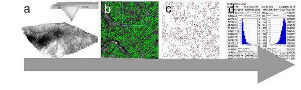
the brandname nAnostic™ (Registered Trademark) and has filed two international patent applications on the process of data extraction from nanoscale cell topographies (WO 2010: PCT/EP2010/057458, PCT/EP2010/068347). The investigation of nanoscale cell topography with AFM is a fairly young discipline (<20 years). A quantification of defined structural elements at the attoliter level (10-18l) using computer vision has been made possible by a proprietary software solution not before last year (Serend-ip). This label free method on data below the optical resolution limits is termed nAnostic™.