
Research
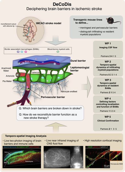
Several barriers at the brain surface and around blood vessels protect the brain from harmful factors from the outside. Breakdown of brain barriers after stroke allows uncontrolled entry of damaging white blood cells and blood components and contributes to brain swelling and damage. Stroke therapies to date have aimed at blocking entry of circulating white blood cells into the brain, with little success. Our approach is different - we will elucidate which of the brain barriers are compromised by ischemic stroke and determine how we can restore the integrity of these barriers after stroke.
We have special genetically modified mice that allow us to see and distinguish the brain barriers, the immune cells that reside at these barriers, and immune cells infiltrating from the blood in the brain of live anesthetized animals. Changes occurring at these barriers as they occur during stroke will be visualized by specialized microscopic techniques, called “intravital microscopy”. Only in this way can factors changing brain barrier properties be identified and validated in human stroke samples.
We expect to identify which of the brain barriers change in function after ischemic stroke. We will further define how these barrier(s) contribute to the entry of potentially damaging immune cells or blood factors after stroke and how their dysfunction contributes to fluid build-up (brain edema). Understanding these mechanisms will permit design of novel therapeutic strategies to stabilize the function of the right barrier after ischemic stroke and, thus, reduce brain damage after stroke and improve the outcome for patients.