

Our Research
We focus on chondrocytes and their surrounding extracellular matrix and evaluate molecular control mechanisms involved in cartilage physiology and pathology.
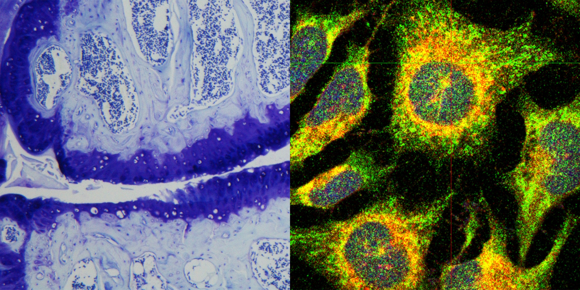
- We are intensively investigating endoplasmic reticulum (ER) stress in chondrocytes, as this stress condition affects viability, activity and differentiation of growth plate and articular chondrocytes and plays a crucial role in the development of chondrodysplasia and osteoarthritis.
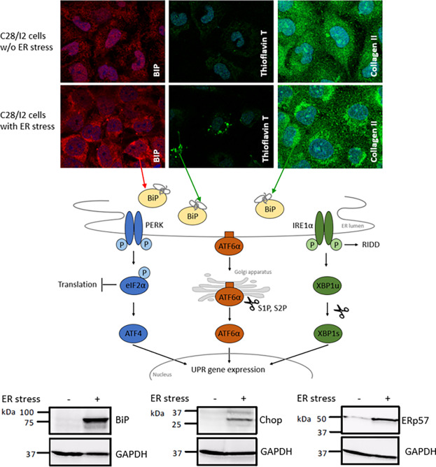
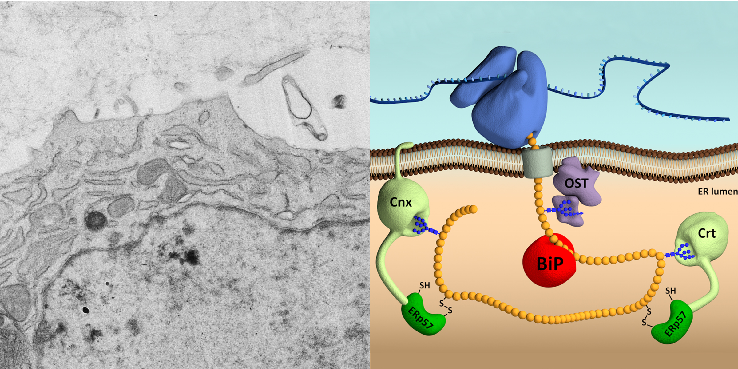
- ER stress in chondrocytes occurs when the capacity of the endoplasmic reticulum to fold proteins is exceeded due to increased levels of secretory proteins, hypoxia, oxidative stress, the presence of mutated proteins to be folded, or loss of functional chaperones, amongst others.
In such situations the unfolded protein response (UPR), an adaptive process to restore normal cell function, is initiated. The UPR triggers reduced translation, increased protein folding, ER biogenesis, augmented degradation of proteins and autophagy. However, if UPR signaling fails to resolve the overload of misfolded proteins in the ER, cell death by apoptosis is induced to remove the stressed cells.
ER stress-induced apoptosis is often associated with loss of cartilage functions in growth plates and joints.
- We are also interested in effects arising from the loss of matrix molecules in cartilage, e.g. decorin or syndecan 4.
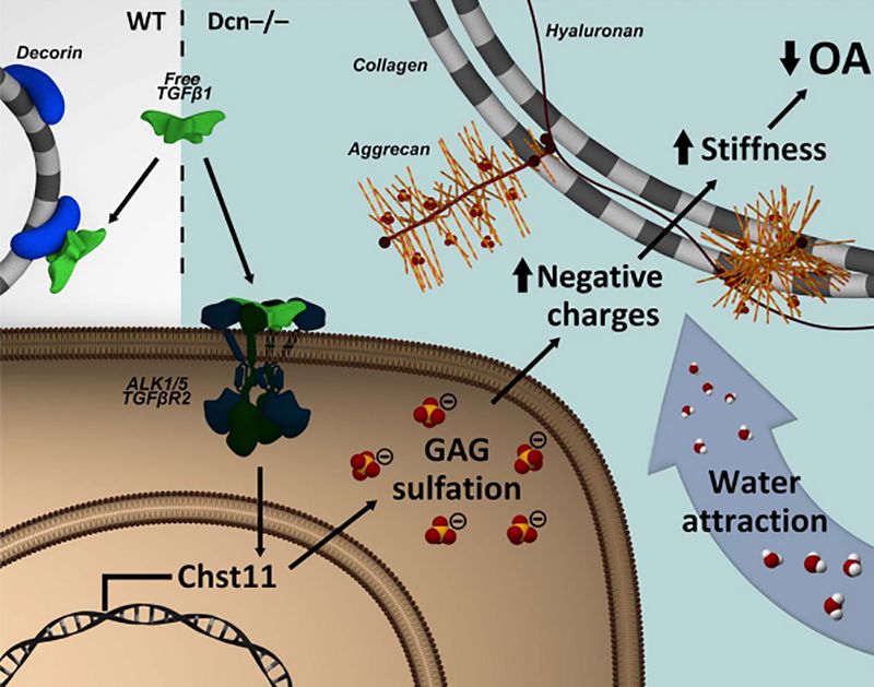
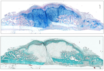
- Interterritorial regions of the articular cartilage matrix are rich in decorin, an important structural protein, also involved in sequestration of TGFβ in the matrix, thereby regulating its activity.
We demonstrated that disruption of decorin-restricted TGFβ signaling leads to higher stiffness of articular cartilage matrix, rendering joints more resistant to OA
- Syndecan 4 is a transmembrane heparan sulfate proteoglycan found in the plasma membrane of chondrocytes.
We have observed that syndecan-4 is dispensable in normal skeletal development since its function is likely to be compensated by syndecan-2. However, in the absence of syndecan 4, bone healing processes were significantly delayed and increased callus formation was observed, suggesting syndecan 4 to be essential during fracture healing.