Frauke Alves

The translation of preclinical findings into clinical application is fundamentally supported by an interdisciplinary working environment and close cooperation with the clinical departments at the University of Göttingen, including the Dept. of Hematology and Oncology (http://www.onkologie-haematologie.med.uni-goettingen.de), the Institute for Diagnostic and Interventional Radiology (http://www.radiologie-umg.de), the Dept. of Nuclear Medicine (http://www.nuklearmedizin.med.uni-goettingen.de), as well as the Heart Centre (http://www.herzzentrum.med.uni-goettingen.de) and the Pulmonary Clinic Immenhausen (http://www.lungenfachklinik-immenhausen.de).
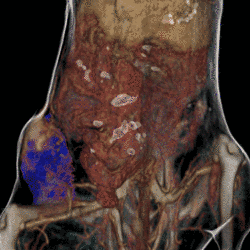
The interdisciplinary group led by F. Alves uses preclinical imaging for various disease mouse models such as for cancer, inflammatory and metabolic diseases. In addition to anatomical imaging, which depicts the structure of disease-related changes in the organism over time, functional procedures such as the optical imaging and SPECT are employed for visualization of biological processes in vivo. The fusion of the two imaging techniques allows the assignment of biological processes to anatomical structures (http://www.imaging-goettingen.de).
Molecular and anatomical imaging is also part of several EU funded projects of the Alves working group, e.g. the project "Nanotechnological toolkits for multi-modal diasease diagnostics and treatment monitoring, NAMDIATREAM" with the aim to develop new nanoparticle-based probes for diagnostics in oncology (http://www.namdiatream.eu), or the Marie Curie Initial Training Network "IonTraC” (IontraC project: Imaging techniques for PDAC diagnostics based on the transportome).
The Industry Academia Partnerships and Pathways Program (IAPP) project entitled "Public-Private Partnership for Asthma Imaging and Genomics, P3AGI", focusing on asthma imaging (http://www.p3agi.eu) and a large collaborative project, "Layered hierarchical structured scaffolds with injectable self setting bioactive gel for clinical bone tissue repair, Innovabone", (https://www.innovabone.eu) are other projects funded by the EU, for which the Alves research team uses non-invasive molecular imaging.
Relevant publications:
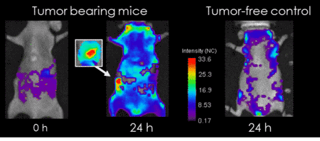
Behnke T, Mathejczyk JE, Brehm R, Würth C, Gomes FR, Dullin C, Napp J, Alves F, Resch-Genger U (2013). Target-specific nanoparticles containing a broad band emissive NIR dye for the sensitive detection and characterization of tumor development. Biomaterials, 34:160-70. Mathejczyk JE, Pauli J, Dullin C, Resch-Genger U, Alves F, Napp J (2012). High-sensitivity detection of breast tumors in vivo by use of a pH-sensitive near-infrared fluorescence probe. J Biomed Opt. Jul;17 Napp J, Behnke T, Fischer L, Würth C, Wottawa M, Katschinski DM, Alves F, Resch-Genger U, Schäferling M (2011). Targeted luminescent near-infrared polymer-nanoprobes for in vivo imaging of tumor hypoxia. Anal Chem, 83: 9039Napp J, Dullin C, Müller F, Uhland K, Petri JB, van de Locht A, Steinmetzer T, Alves F (2010). Time-domain in vivo near infrared fluorescence imaging for evaluation of matriptase as a potential target for the development of novel, inhibitor-based tumor therapies. Int J Cancer. 127:1958-74. Dullin C, Zientkowska M, Napp J, Missbach-Guentner J, Krell HW, Müller F, Grabbe E, Tietze LF, Alves F (2009). Semiautomatic landmark-based two-dimensional-three-dimensional image fusion in living mice: correlation of near-infrared fluorescence imaging of Cy5.5-labeled antibodies with flat-panel volume computed tomography. Mol Imaging; 8:2-14.
Behnke T, Mathejczyk JE, Brehm R, Würth C, Gomes FR, Dullin C, Napp J, Alves F, Resch-Genger U (2013). Target-specific nanoparticles containing a broad band emissive NIR dye for the sensitive detection and characterization of tumor development. Biomaterials, 34:160-70. Mathejczyk JE, Pauli J, Dullin C, Resch-Genger U, Alves F, Napp J (2012). High-sensitivity detection of breast tumors in vivo by use of a pH-sensitive near-infrared fluorescence probe. J Biomed Opt. Jul;17 Napp J, Behnke T, Fischer L, Würth C, Wottawa M, Katschinski DM, Alves F, Resch-Genger U, Schäferling M (2011). Targeted luminescent near-infrared polymer-nanoprobes for in vivo imaging of tumor hypoxia. Anal Chem, 83: 9039Napp J, Dullin C, Müller F, Uhland K, Petri JB, van de Locht A, Steinmetzer T, Alves F (2010). Time-domain in vivo near infrared fluorescence imaging for evaluation of matriptase as a potential target for the development of novel, inhibitor-based tumor therapies. Int J Cancer. 127:1958-74. Dullin C, Zientkowska M, Napp J, Missbach-Guentner J, Krell HW, Müller F, Grabbe E, Tietze LF, Alves F (2009). Semiautomatic landmark-based two-dimensional-three-dimensional image fusion in living mice: correlation of near-infrared fluorescence imaging of Cy5.5-labeled antibodies with flat-panel volume computed tomography. Mol Imaging; 8:2-14.