
News
Das aktuelle "Paper of the Month" (05/2018) geht an: Priv.-Doz. Dr. Astrid Jeibmann aus dem Institut für Neuropathologie
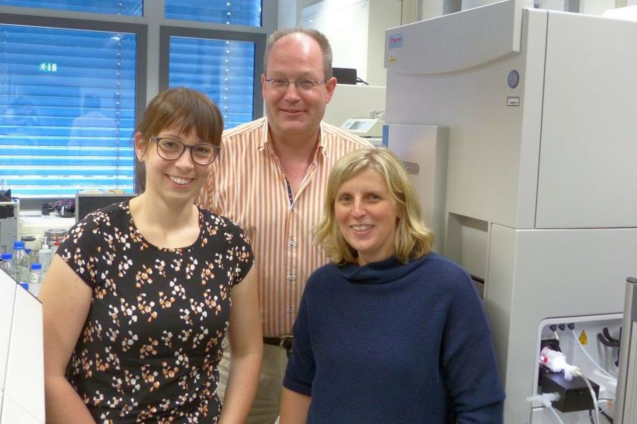
PD Dr. Astrid Jeibmann (r., geteilte Letztautorenschaft) mit Prof. Uwe Karst (m., dito) und Stefanie Fingerhut (Erstautorin) (Foto: Sabrina Funke)
Für den Monat Mai 2018 geht das "Paper of the Month" der Medizinischen Fakultät der WWU Münster an:
PD Dr. Astrid Jeibmann aus dem Institut für Neuropathologie für die Publikation:
Zu Hintergrund, Fragestellung und Bedeutung der Publikation:
Kürzlich veröffentlichte Studien konnten zeigen, dass nach mehrmaliger Gabe von Gadolinium-haltigen Kontrastmitteln im Rahmen der MRT-Bildgebung Gadolinium-Ablagerungen in verschiedenen Bereichen des Gehirns auftraten, am stärksten sichtbar im Nucleus dentatus des Kleinhirns. Auch wenn keine klinischen Korrelate bisher bekannt sind, gibt es eine intensive Debatte, ob diese Ablagerungen schädlich sein können.
Das Ziel unserer Studie war es, die Gadolinium-Verteilung im Gehirngewebe von Patienten, die mehrfache Injektionen Gadolinium-haltiger Kontrastmittel erhielten, im niedrigen Mikrometerbereich zu untersuchen und mögliche pathologische Gewebeveränderungen durch Gadolinium-Ablagerungen zu untersuchen. Wir konnten mit einer analytischen Methode, die die Untersuchung ortsaufgelöster Elementverteilungen ermöglicht (LA-ICP-MS) Gadolinium-Ablagerungen in den Gefäßwänden kleiner Blutgefäße im Nucleus dentatus bei allen Patienten nachweisen, die Gadolinium-haltige Kontrastmittel erhalten haben, während wir diese in der Kontrollgruppe nicht nachweisen konnten. Es konnten bei keinem der Patienten signifikante pathologischen Veränderungen des Gehirngewebes gefunden werden.
Wir konnten zeigen, dass Gadolinium-Ablagerungen im Gehirn auf die Blutgefäßwände beschränkt sind, das Neuropil nicht betroffen ist und Reaktionen auf zellulärer Ebene nicht nachweisbar sind.
Background and fundamental question of the publication:
Recent studies showed gadolinium depositions following serial administrations of gadolinium-based contrast agents (GBCAs) for magnetic resonance imaging examinations in various parts of the brain with the dentate nucleus being most affected. Even though no clinical correlates of the deposits were known yet, an intensive debate developed if this might be harmful.
The aim of the current study was to specify the gadolinium distribution in brain tissue of patients who received serial injections of GBCAs in the low-µm range and to explore any potential pathological tissue changes caused by gadolinium deposits. Using an analytical method, allowing for spatially resolved detection of chemical elements (LA-ICP-MS) revealed gadolinium depositions in the walls of small blood vessels of the dentate nucleus in all GBCA exposed patients, while no gadolinium was found in the control group. No significant pathological changes of the brain tissue with respect to micro-/astrogliosis and neuronal loss were found in any of the patients.
The findings show that gadolinium depositions in the brain are restricted to blood vessel walls, while the neuropil is spared and apparent cellular reactions are absent.
Die bisherigen ausgezeichneten "Papers of the Month" finden Sie HIER.
This could be interesting for you too:
Für den Monat Dezember 2019 geht das "Paper of the Month" der Medizinischen Fakultät der WWU Münster an: Dr. Max Masthoff aus dem Institut für Klinische Radiologie für…
Münster (mfm/tw) - Reisen zum Mittelpunkt der Zelle: Ein neues Mikroskop am Zentrum für Pathologie, Anatomie und Neuropathologie (PAN-Zentrum) der Medizinischen Fakultät der Universität…
Münster (mfm/tb) - Wie tief ist der Tumor bereits in das Gewebe eingedrungen? Um Patienten mit einem Hirntumor wirksam therapieren zu können, muss sich der behandelnde Arzt ein möglichst…