Reference of fragmentation data of single amino acids prepared by electrosprayIn order to understand mass spectra of unknown substances it is sometimes necessary to analyse synthetic standards. Below is a collection of MS/MS spectra taken with either LCQ or TSQ-700 from Finnigan MAT (for details see S. Königs & H. M. Fales (2011) Biomacromol. Mass Spectrom. 2,3: 211-220). The data were first shown at the 48th Conference of the American Society for Mass Spectrometry in Long Beach, CA.
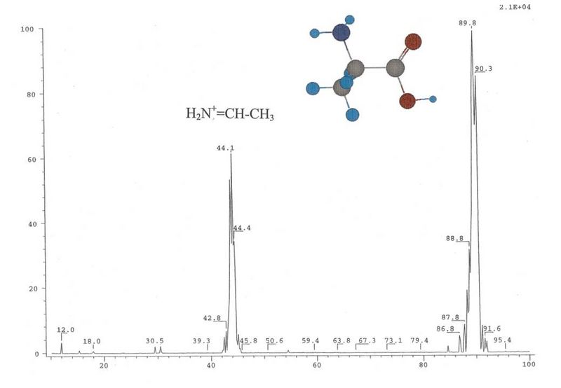
Alanine
TSQ MS/MS at -20 eV. No ammonia and water losses are abserved.
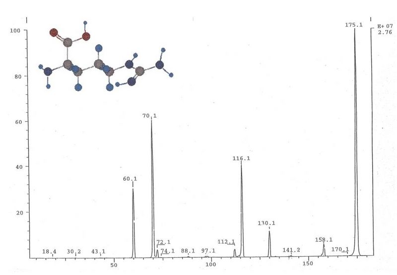
Arginine
TSQ MS/MS at -20 eV. Arg first loses either 17 or 45 Da and only then the usual 18 and 46 Da as seen in the TSQ. However, in the ion trap a primary water loss is observed while formic acid is lost only after the removal of three terminal nitrogen atoms from both ends of the molecule.
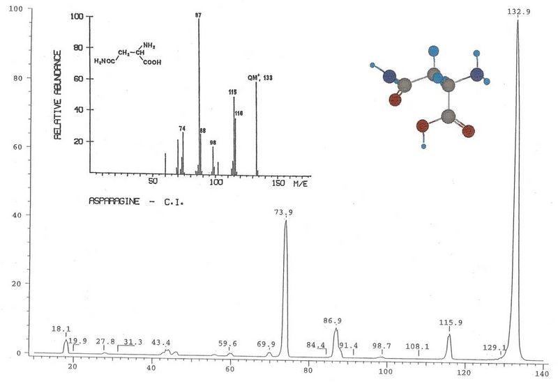
Asparagine
The ESI spectrum (TSQ MS/MS, -20 eV, in comparison to CI spectrum) resembles in all major features the CI spectrum. It is characterized by two losses of ammonia and the loss of formic acid as well as the loss of the side chain as CH3-CO-NH2. The loss of 45 Da from the parent ion as suspected from the TSQ data cannot be confirmed in the ion trap. As expected, aspartic acid behaves in much the same way, differing only in the fact that it loses water instead of ammonia and, accordingly, acetic acid.
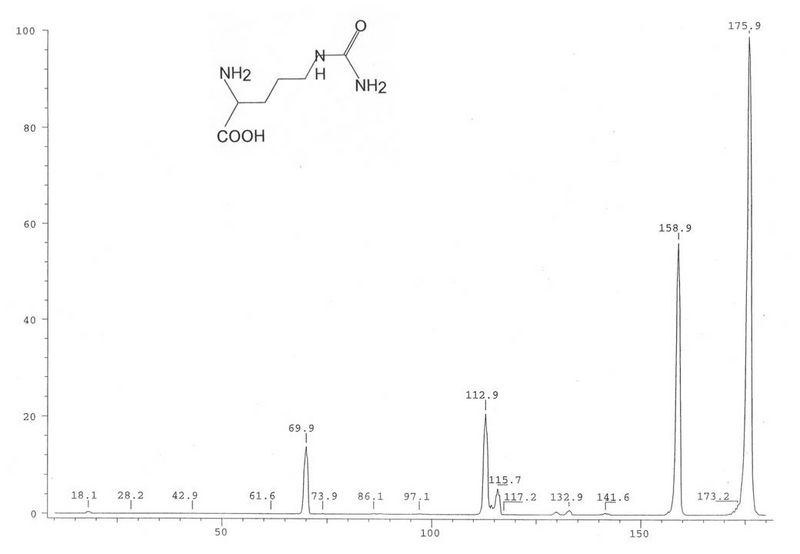
Citrulline and ornithine spectra are characterized not only by the ususal losses of ammonia and formic acid but also by the loss of formamide which, in the case of citrulline, is already evident in the TSQ spectrum. It appears in the ion trap only in MS3 of m/z 115 following the water loss. For citrulline, the loss of formamide follows the loss of 17 and 43 Da suggesting ring formation for m/z 116. Histidine is the only basic amino acid which shows a HCN loss following the removal of HCOOH which may be due to the formation of a six-membered ring structure.
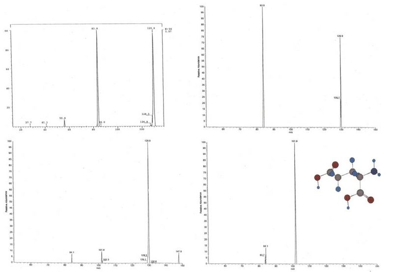
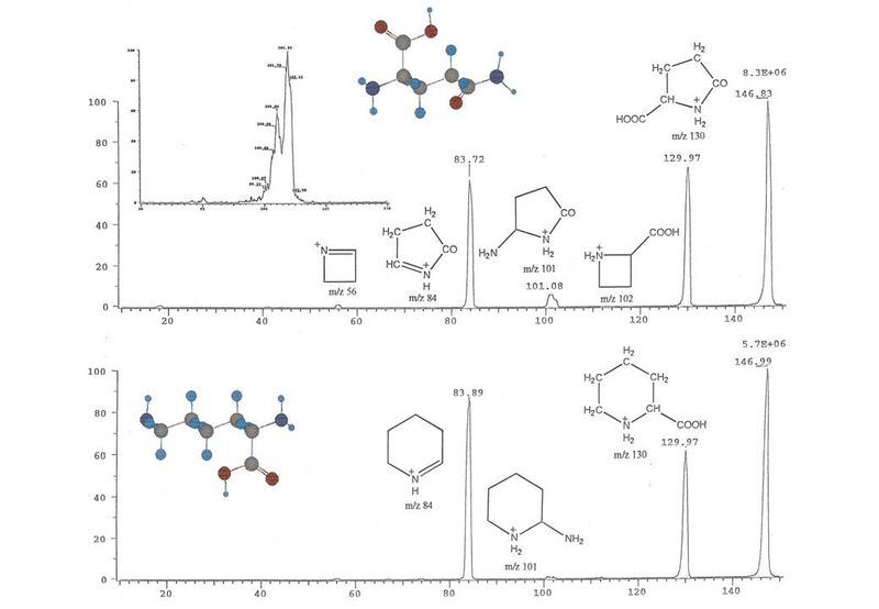
Glutamic acid
Upper left: TSQ MS/MS of pyrogutamic acid at -29 eV.Lower left: LCQ MS/MS of Glu at 9% collision energy.Upper right: MS3 of m/z 130 at 9.9%.Lower left: MS3 of m/z 102 at 9.9%.Pyroglutamic acid is a dominant fragment and in turn can lose the elements of formic acid to form the fragment at m/z 84.
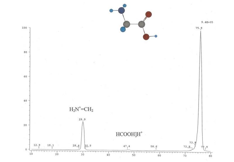
Glycine
TSQ MS/MS at -20 eV.
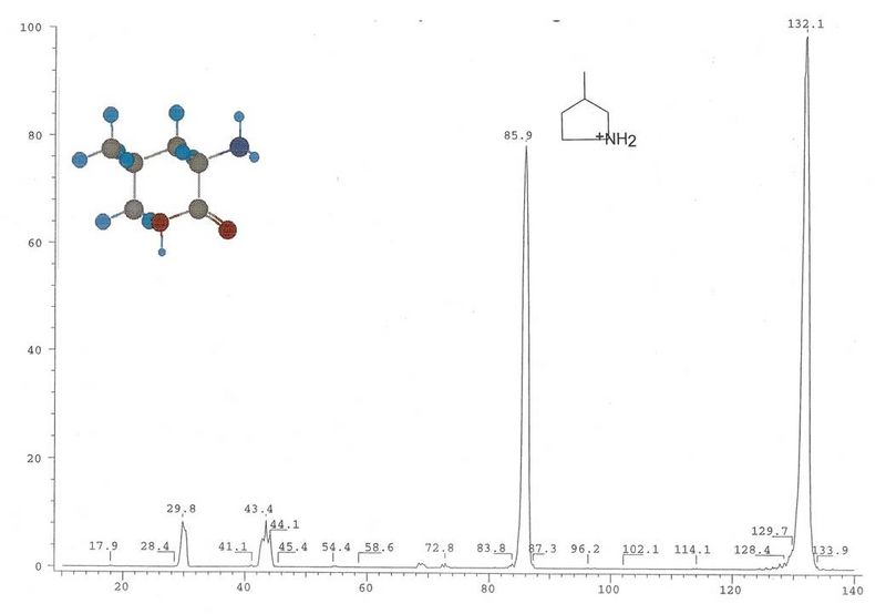
Isoleucine
Leucine
TSQ MS/MS at -20 eV. Does not break further than m/z 86 in LCQ possibly due to ring formation.
Lysine - Glutamine
MS/MS spectra at -15 eV collision energy. The high resolution scan of the ions at m/z 101/102 shown in the inset of the Gln-spectrum (top trace; scan m/z 90-110 in 1 min, at 576 samples per peak, daughter resolution increased by 5 nominal units, acquisition time 3 min) shows the fragment ions resulting from the loss of 46 and 45 da from the parent ion. A weak peak can be observed at about 1% at m/z 101 for Lys. For discussion see paper.
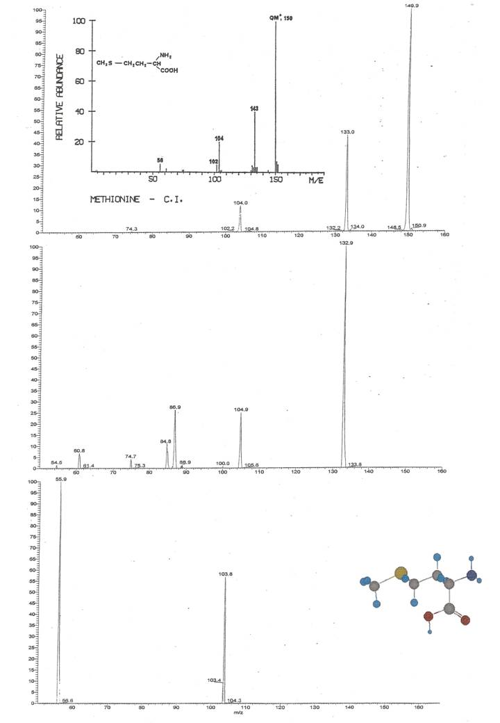
Methionine
LCQ MSn in comparison to CI. MS2 of m/z 150 at 8%. MS2 of m/z 133 at 10%. MS3 of m/z 104 at 8%.As shows best in the ion trap, methionine preferably loses either formic acid or ammonia rather than water first. As revealed in MS3 of m/z 133, the loss of 17 Da follow losses of either 46 Da, or 48 Da for methy thiol, and protonated acetic or propionic acid are present at m/z 61 and 75, respectively. MS3 of m/z 104 shows that only the loss of 48 Da (CH3-SH) is present at collision energies of 8%.Cysteine was present as dimer; fragmentation of the monomer was difficult to achieve without previous reduction. The dimer fragments to retain buth sulfor atoms on the ion at m/z 152, and goes then on to lose first one sulfur to form the thiol and then the elements of formic acid as seen at -30 eV in the TSQ. The spectrum shows more complexity in th eion tap at 12% collision enegry and smaller losses such as 17, 18, 26, and 46 Da are more pronounced.
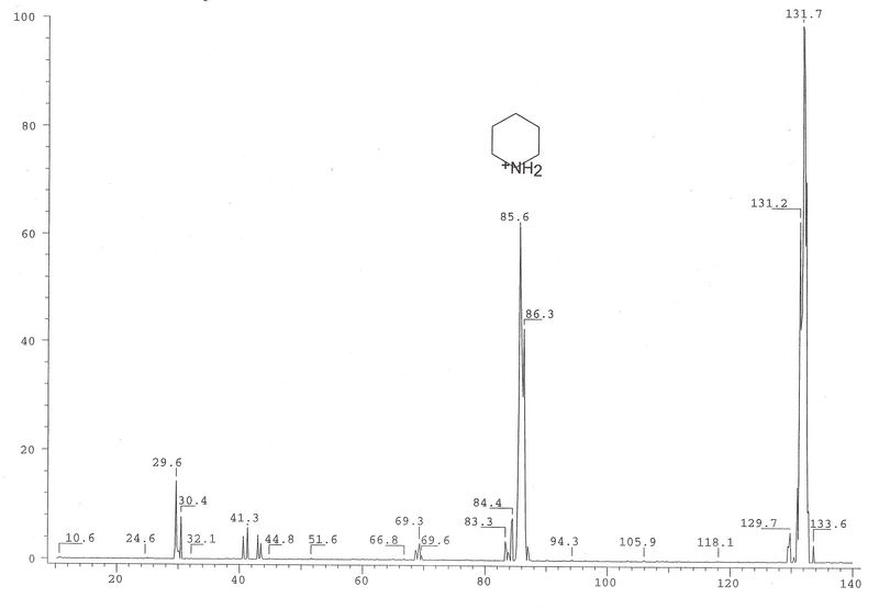
Norleucine
TSQ MS/MS at -20 eV. The 17 loss from m/z 86 has been observed in the LCQ.
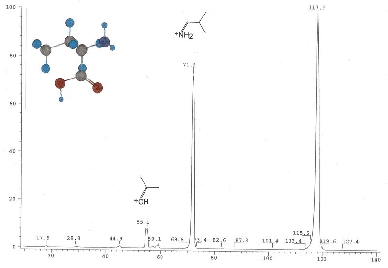
Norvaline
Possible ring formation stabilizes m/z 72, therefore, less fragments at m/z 55, 57, 59 are found, but more intense m/z 30 as in valine.
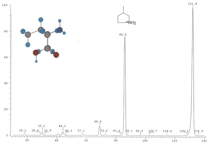
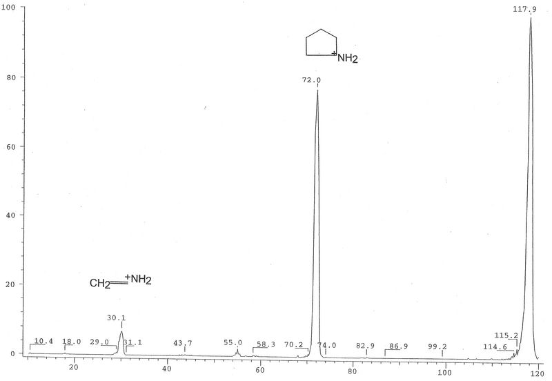
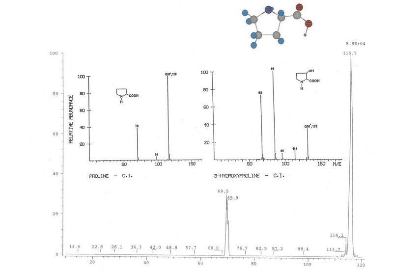
Proline
TQS MS/MS at -15 eV. No water loss is observed. Inset CI data.
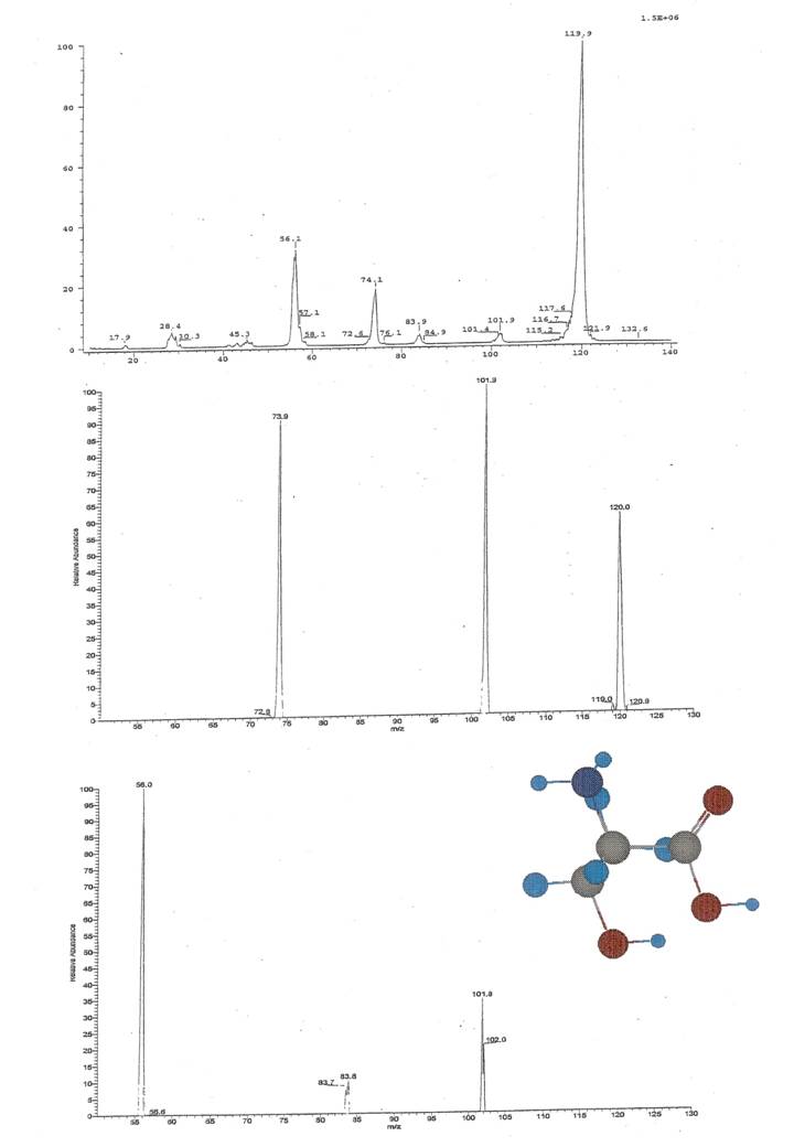
Threonine
Top: TSQ MS/MS at -30 eV.Middle: LCQ MS/MS at 9%.Bottom: LCQ MS3 of m/z 102 at 9.9%.As can be expected from the presence of two hydroxyl groups in the molecules, both Ser and Thr successively lose two molecules of water, or water and formic acid in either order. In MS/MS in the ion trap only the water loss from the C-terminus or the loss of HCOOH is observed at 9% collision energy while the second water loss is seen in MS3. No ammonia loss can be found in the ion trap while both compounds show some minor loss of 17 Da following the loss of 46 Da.