Ultrastructural analysis using electron microscopical methods
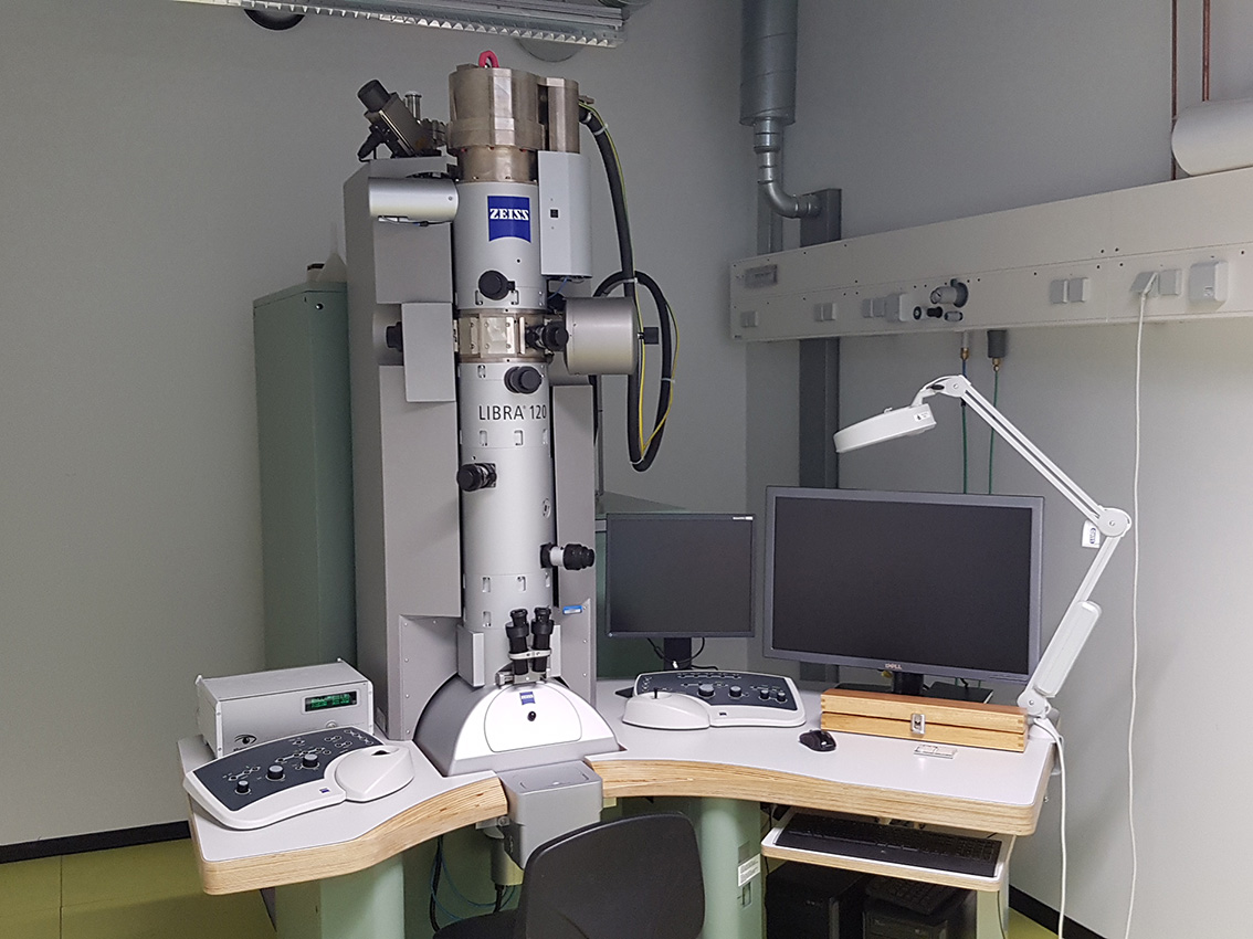
Transmission-Electron Microscope (TEM) " Zeiss LIBRA® 120 " with optional GATAN Cryo Unit
Applications: » conventional TEM and Cryo-EM ( no tomography )Additional equipment: » Leica High Pressure Freezer "EM PACT2" » Leica Automatic Freeze Substitution System "EM AFS2" » Leica Ultracut "EM UC7" inc. Cryosphere Contact: Dr. Astrid Rohlmann Tel.: 0251 83-50207 Astrid Rohlmann
Our group analyzes neuronal synapses of different genetically modified mouse lines in vivo and in vitro biochemically, electrophysiologically and morphometrically. For the latter, light microscopical techniques and hence electronmicroscopical experiments are performed. Morphometrical analysis with respect to questions of number, ultrastructural parameters etc. of synapses is done at the transmission electron microscope (TEM) with appropriate software.
Different techniques for fixation of mouse brains and shock freezing of native brains using high pressure freezing (HPF) and subsequent automatic freeze substitution (AFS), as well as different pre- or postembedding immunolabelings (immunoEM) are established.
We are open to discuss putative TEM / immunoEM projects with other labs on a collaborative basis.