
Service
Sample preparation for transmission electron microscopy
- Sample preparation for cryo-immobilization (High-pressure freezing, Plunge freezing, freeze fracture, replica), or chemical fixation
- Sectioning: ultrathin, semi-thin, serial sections
- Positive contrasting (pre- or post-embedding) and negative staining
- Immuno-labelling/staining (pre-embedding or labeling on plastic sections)
- Grid preparation
Sample preparation for scanning electron microscopy
- Chemical fixation
- critical point drying
- Freeze drying
- Cryo-immobilization (High-pressure freezing, Plunge freezing, freeze fracturing)
- Cryo, high-vacuum transfer
- Conductive coating (Sputter and Evaporation)
Facility
- Provision of EM-specified chemicals and supplies for users
- Support and training in the methods listed in “Service” above
- Access to the equipment of the facility
- Training and support in ultramicrotome and electron microscope usage
- Support and assistance in cell morphology, ultrastructure and data interpretation
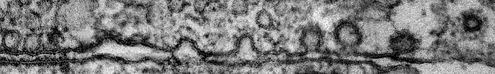
Imaging
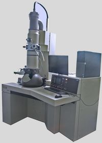
Transmission electron microscope Phillips CM10
This TEM can operate with different accelerating voltages (40-100 kV in 20 kV steps) and achieves the resolution up to 0.5 nm thus being suitable for a variety of biological as well as other types of samples. Microprocessor-controlled operation unit allows easy and stable usage of the microscope. High-contrast objective lens of this instrument together with bottom-mounted dedicated high-resolution EM camera TEMCam F-416 from TVIPS provide excellent images in every-day use. Original image acquisition software from TVIPS allows for an easy operation.
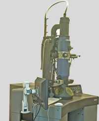
Transmission electron microscope Phillips 410
With 0.4 nm resolution at 100 kV accelerating voltage combined with 5 Mpix side-mounted CMOS camera Phillips 410 TEM is perfectly suited for routine examination of variety of samples. Additionally there is a possibility to use a bottom-mounted plate’s camera with high-resolution Imaging Plates (Ditabis).
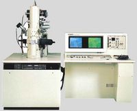
Scanning electron microscope Hitachi S5000
High-resolution In-Lens Cold-Field Emission SEM achieves the resolution up to 0.6 nm (at accelerating voltage 30kV). Apart from SE- and BSE-detectors it is also equipped with STEM-module. The STEM-detector allows acquisition of high-resolution transmission images from not contrasted samples along with the mass determination. For the examination of biological samples at low temperature Oxford Cryoholder CT3500 can be used. Freeze-drying system together with cryo-vacuum-transfer unit were custom developed in our facility to extend the examination capabilities of frozen biological samples. The active picture acquisition and analysis system for scanning electron microscopes (DISS5; point electronic) allows simultaneous acquisition of signals from all detectors as well as on-line analysis.
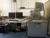
Scanning electron microscope Hitachi S4700
High-resolution Field Emission SEM achieves the resolution up to 1,5 nm at 15 kV and 2,5 nm at 1 kV. Apart from SE- and BSE-detectors it is also equipped with In-Lens SE-detector for HR-mode. Routine microscopy at working distance of 12 mm. Motorized 5 axis sample stage (100x50 mm). Fully mouse or keyboard operated PC controlled instrument. Built-in Anticontaminator. The active picture acquisition and analysis system for scanning electron microscopes (DISS5; point electronic) allows simultaneous acquisition of signals from all detectors as well as on-line analysis.
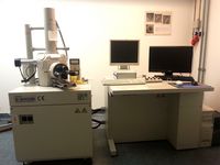
Scanning electron microscope Hitachi S3000N
Universal computer controlled scanning electron microscope with resolution of 3.5 nm and big 150 mm specimen chamber can provide a quick overview of a SEM sample. High sensitivity Quadrant Type Semiconductor BSE Detector. It is equipped with digital image acquisition system (DISS5; point electronic).
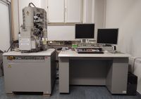
Scanning electron microscope FEI Quanta 600F incl. Leica cryo stage
The Quanta scanning electron microscope is a versatile, high-performance instrument with three modes (high vacuum, low vacuum and ESEM) to accommodate the widest range of samples. The field emission gun system is equipped with EDX analysis and motorized 150 mm stage. Samples can be investigated at cryo temperature via high vacuum cryo transfer system Leica VCT500 and cryo stage
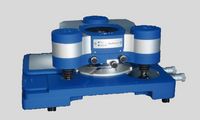
Atomic force microscope JPK NanoWizard II
NanoWizard® II provides the highest resolution and stability and appeared to be the leading solution for the applications which would require the integration of modern optical methods with highest efficiency of AFM. The area of use includes mapping of living cells, experiments on single molecule or nano-particle level in biophysics, pharmacology and cellular biology. It utilizes also the patented DirectOverlay™-function which allows the direct combining of AFM- and optical microscopy images distortion free. (For more information please follow the link: http://www.jpk.com/nanowizard-r-ii-overview.351.en.html)
Sample preparation equipment
There is a variety of different instruments used for the sample preparation either for SEM or TEM. This includes:
- Fresh and fixed tissue preparation: vibrating blade microtome Leica VT1000 S
- Leica EM ACE900 freeze fracture system
- Room-temperature ultramicrotomes (Reichert, Reichert Cryo, Leica Ultracut EM UC7)
- High-pressure-freezing machine HPM-100 with optical stimulation (Leica)
- Freeze-substitution machine AFS-2 (Leica)
- Plunge freezer
- Sputter coater
- Thermal and electron beam evaporators
- High vacuum cryo shuttle
- Critical-point dryer
- Freeze-drying machines